SAN FRANCISCO - New clinical trial results show that the presence of vulnerable plaque on VH intravascular ultrasound (IVUS) imaging before percutaneous coronary intervention (PCI) is significantly associated with poorer three-year outcomes.
Conversely, the absence of at least a 10% necrotic core at baseline on VH IVUS (Volcano Therapeutics, San Diego, CA) imaging was a strong predictor of lesion stability, according to lead investigator Dr. Gregg Stone from Columbia University Medical Center In New York City.
"Looking at the angiographic severity of the blockage is not enough … we must understand the type of disease present in the vessel wall," Stone said.
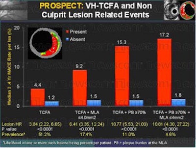
The PROSPECT (Providing Regional Observations to Study Predictors of Events in the Coronary Tree) trial examined 700 patients at 40 clinical centers in the U.S. and Europe. Following VH IVUS imaging of the target vessels, all patients received PCI for acute coronary syndromes (ACS) including unstable angina, ST-segment elevation myocardial infarction (STEMI), or non-T-segment elevation myocardial infarction (NSTEMI).
The hazard ratio for a thin-cap fibroatheroma (TCFA) identified at VH IVUS was 3.8 (p < 0.0001), indicating a statistically significant predictor of an event, versus a hazard ratio of 0.2 for pathological intimal thickening (PIT) lesions, which were significantly less likely to cause an event.
The absence of a TCFA (< 10% necrotic core at VH IVUS) was a strong predictor of lesion stability over a three-year follow-up period. The likelihood of the lesion progressing to an event over three years was negligible, independent of other grayscale and angiographic factors, the researchers reported.
The plaque type yielding the highest risks were fibroatheromas with a minimum lumen area of ≤ 4 mm2 and a plaque burden ≥ 70%. Three independent predictors combined showed that such a lesion imaged at baseline had a 17.2% chance of causing an adverse cardiac event within three years.
 |
| Lesions consisting of >10% thin-cap fibrous atheroma were far likelier to cause adverse events within three years of percutaneous coronary intervention. Image courtesy of Volcano Therapeutics. |
The study demonstrates that lesions characterized as mild on angiography are capable of progressing quickly to cause events, and that the sites of adverse events are often missed at angiography, according to Volcano. Current practice using angiography guidance alone is insufficient in that regard, the company added.
Related Reading
Plaque morphology imaging improves PCI outcomes, September 25, 2009
Primary PCI vs. prehospital lysis for MI: similar survival, August 19, 2009
Post-PCI eptifibatide infusion can be safely cut from 18 to 2 hours, March 12, 2009
Percutaneous cardiovascular procedures 'strikingly better' with radial access, March 10, 2009
CABG remains superior to PCI for severe coronary disease, February 19, 2009
Copyright © 2009 AuntMinnie.com