A new method for segmenting lung nodules in 3D CT images shows promise for improving the quantification, detection, and diagnosis of nodules, according to research from the University of Chicago in Illinois.
"The experiments demonstrated that our segmentation method provided quite robust and accurate results," said Jiahui Wang, Ph.D.
As segmentation of lung nodules is one of the most important steps in a computer-aided detection (CAD) scheme, the Chicago researchers sought to develop a reliable method of segmenting lung nodules in 3D images using what they call a "spiral scanning" technique.
After determining a volume of interest (VOI) at the nodule location, the 3D VOI is transformed into 2D via the spiral scanning technique. In this method, a radial line originating from the center of the VOI spirally scans the VOI from its north pole to its south pole, Wang said. Now at Duke University in Durham, NC, he presented the research at the 2007 RSNA meeting in Chicago.
Segmentation is then performed on the resulting 2D image using dynamic programming, which involves searching for end points of an optimal outline, column by column, according to Wang. The result is a continuous and smooth outline, he said.
Next, the 2D image is transformed back into the 3D image by transforming points on a 2D outline to points on a 3D surface. The 3D surface is then filled to obtain a solid volume, according to Wang.
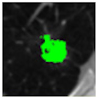
 |
| Image courtesy of Jiahui Wang, Ph.D. |
To test its technique, the study team made use of a CT database provided by the Lung Imaging Database Consortium (LIDC). The database included two LIDC datasets; the first dataset included 23 nodules with a size range of 4.0 mm to 33.6 mm and a slice thickness ranging from 0.6 mm to 2.5 mm. The second dataset had 73 nodules with a size range of 3.8 mm to 30.2 mm and slice thickness of 1.3 mm to 3.0 mm.
The researchers trained the method on the first dataset, then evaluated it on the second. Four to six chest radiologists had provided the outlines for each nodule to serve as the gold standard.
The study team evaluated the performance of the segmentation method by examining the overlap value between the computer segmentation and the gold standard. The segmentation method produced a mean overlap value of 66% for the 23 training nodules, and 64% for the 73 testing nodules. In comparison, researchers at two other institutions had previously provided overlap values of 58% and 51% for the 23 nodules in the first LIDC dataset.
The researchers found higher accuracy for larger nodules than for smaller ones. The mean overlap values were 63% for the 52 small (< 10 mm) nodules and 70% for larger nodules.
"A key contribution is the spiral scanning technique, which considerably simplified the segmentation scheme and improved the segmentation performance," Wang said.
By Erik L. Ridley
AuntMinnie.com staff writer
February 28, 2008
Related Reading
Hybrid lung segmentation software boosts performance, February 13, 2008
Experience may make a difference with lung CAD, February 4, 2008
Radiologists more likely to reject certain true-positive CAD findings, January 22, 2008
CAD offers value in detecting lung nodules with CT, December 17, 2007
CAD may boost radiologists' ability to characterize lung nodules, November 25, 2007
Copyright © 2008 AuntMinnie.com