The search for an alternative to mammography continues with two recent studies that evaluated the role of CT in breast imaging. In the first, a Houston-based group sought to determine how conebeam CT fared for breast tissue contrast and overall exam results. In the second, California-based investigators updated their ongoing studies with the modality, looking specifically at improving image quality.
True 3D
Based on their results, Dr. Wei Tse Yang and colleagues suggested that dedicated conebeam breast CT showed particular promise for diagnosing tumors at an early stage because it can overcome the problem of overlapping tissue through "true 3D" acquisition.
Yang's group is from the departments of radiology, imaging physics, and pathology at the M. D. Anderson Cancer Center, University of Texas, in Houston.
For their research, they imaged 12 mastectomy specimens on an experimental conebeam CT scanner with an amorphous silicon flat-panel digital detector (Paxscan 4030CB, Varian Medical Systems, Palo Alto, CA). Scanning was done at 50-80 kVp with reconstructed voxel size of 145 µm or 290 µm. The calculated dose levels were equivalent to 6-25 mGy for one breast, corresponding to one to four times the mean glandular dose limits for two-view mammograms of a 5-cm thick compressed breast.
According to the results, the mean scanning time was 12 seconds for images that were deemed adequate for tissue visualization and 48 seconds for high-resolution images. On the latter, cancer appeared as an "irregular spiculated mass with associated microcalcifications or overlying skin thickening, or an area of focal asymmetry."
Microcalcifications within cancers were clearly seen with conebeam CT imaging (American Journal of Roentgenology, December 2007, Vol. 189:6, pp. 1312-1315).
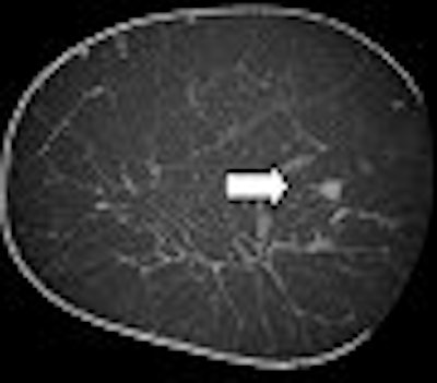
 |
| A 63-year-old woman with invasive ductal carcinoma of left breast. Coronal CT image shows microcalcifications within area of architectural distortion representing known cancer (arrows). Pathology showed ductal carcinoma in situ associated with microcalcifications. Yang WT, Carkaci S, Chen L, Lai C, Sahin A, Whitman GJ, and Shaw CC, "Dedicated Cone-Beam Breast CT: Feasibility Study with Surgical Mastectomy Specimens" (AJR 2007; 189:1312-1315). |
The group also noted that conebeam CT may be particularly useful for imaging the postoperative breast, which can be hampered by overlapping structures, skin thickening, and simulation of pseudomasses. Other potential uses would be for image-guided surgery and interventional procedures.
"The advantages of conebeam CT include ... true 3D images with isotropic resolution, reduced motion artifacts, breast-only exposure to radiation ... and high contrast resolution," the researchers concluded. "Further study of conebeam CT with larger series is needed to better understand its potential role in ... breast cancers."
Technical success
At the 2005 RSNA meeting in Chicago, California-based researchers discussed their work with a prototype conebeam CT scanner. At the time, John Boone, Ph.D.; Dr. Karen Lindfors; and colleagues had conducted a preliminary study of 21 prebiopsy patients who were BI-RADS 4 or 5 on mammography. Boone and Lindfors are from the Sacramento-based medical center of the University of California, Davis (UCD).
According to their 2005 subjective results, lesions detected on mammography were also seen on breast CT in 19 of the 21 cases. Another benefit of the CT exam was self-reported comfort by the patients because no compression was required. However, there were issues with spatial resolution with slight image degradation at the periphery of the images.
 |
| Breast CT shows an ill-defined mass (ductal carcinoma in situ [DCIS]). Image courtesy of John Boone, Ph.D. |
At the 2007 RSNA meeting, the group focused on a poster presentation on image quality from scans done on 103 women on two prototype scanners, also outfitted with amorphous silicon flat-panel digital detectors (Paxscan 4030CB). Boone is a research consultant for Varian Medical Systems.
In this study, they measured image quality by looking at modulation transfer function (MTF), noise power spectrum (NPS) parameters, and root mean score (RMS) noise measurements. "Images from each breast compromise 300 to 500 images, each with 512 x 512 voxels measuring about 0.30 mm isotropically," they explained in their RSNA abstract.
 |
| A 4-mm intraductal carcinoma (IDC) on mammography (above) and breast CT (below). Image courtesy of John Boone, Ph.D. |
 |
The researchers found that the measured MTF was superior to conventional whole-body CT (Fc about 2 cycles/mm). While the NPS level was slightly higher, the appropriate choice of section thickness makes the breast CT images acceptable, they added.
"There is a need for higher resolution in breast imaging because some early warning signs of cancer are very small microcalcifications," wrote Boone in an e-mail to AuntMinnie.com, explaining the significance of these MTF results.
 |
| A 7-mm cluster of suspicious microcalcifications. Coronal and axial breast CT scans show microcalcifications in an ill-defined mass (DCIS). Image courtesy of John Boone, Ph.D. |
"That is why mammography has far better resolution than any other radiography procedure performed.... The better MTF reflects the need to design the breast CT scanner so that it has spatial resolution that is as good as possible," added Boone, who is the vice chair of radiology research and a professor of radiology and biomedical engineering at UCD.
To date, Boone and co-investigators have performed breast CT exams in 139 patients and are continuing to accrue patients as part of a phase II trial. In the meantime, the group just published more data from its study, detailing breast CT results in 69 women.
These results echoed those from another recent study, with CT turning in a performance that was equal to mammography for visualization of breast lesions, particularly masses. The researchers also did not see significant differences between CT and mammography results based on breast density. Finally, the subjects found CT more comfortable than mammography once again (Radiology, January 14, 2008).
By Shalmali Pal
AuntMinnie.com staff writer
January 25, 2008
Related Reading
Imaging start-up to install conebeam CT breast imaging prototype, May 1, 2006
Breast CT developers aim to 'revolutionize' mammography, December 20, 2005
Researchers make inroads with breast CT, gear up for clinical test, July 25, 2005
Copyright © 2008 AuntMinnie.com