Volumetric and multiplanar-reformatted (MPR) CT yielded significantly better results than transverse views for gastric cancer staging, researchers from Korea have reported. The volumetric advantage wasn't statistically significant for detecting metastases and lymph nodes, however.
"Although clinically important advances in diagnosis, such as endoscopy and double-contrast barium swallow studies, allow the detection of small lesions early in the course of the disease, the depth of tumor invasion and the presence or absences of metastases cannot be determined with these modalities," wrote Dr. Hye Jin Kim, Dr. Ah Young Kim, and colleagues from the University of Ulsan College of Medicine in Seoul (Radiology, September 2005, Vol. 236:3, pp. 879-885).
This task is usually delegated to thin-slice multidetector-row CT. And while the authors noted that it is "natural to suspect" that CT gastrography performed with MPR and a virtual endoscopic technique could be helpful, "to our knowledge there has been minimal focus on the diagnostic value of volumetric CT imaging of patients with gastric cancer," they wrote.
To address this issue, the study team examined 106 patients (102 men, 34 women; mean age 56 years) with endoscopically proven gastric cancer, prior to surgery and within five to 12 days following endoscopic biopsy.
Forty-five patients had early-stage cancer and 61 had late-stage disease. Pathologic TNM stage was defined as pT1, tumor invasion of the mucosa and/or mulscularis mucosa or submucosa; pT2, tumor invasion o the muscularis propria or subserosa; pT3, tumor penetration of serosa; and pT4, tumor invasion of adjacent structures.
Lymph nodes were categorized as N0 for no evidence of lymph node metastasis; N1, metastasis to group 1 lymph nodes; N2, metastasis to group 2 lymph nodes; and N3, metastasis to group 3 lymph nodes. Metastasis was classified as M0 or M1, depending on whether distal metastases were observed, the authors wrote.
After fasting to empty the stomach, 20 mg of scopolamine was administered (Buscopan, Boehringer Ingelheim, Ingelheim, Germany) in all patients, and CT images were acquired using a four-slice scanner (LightSpeed QX/i, GE Healthcare, Chalfont St. Giles, U.K.).
The scan parameters included 120 kVp, 200 mAs, 1.5-mm collimation, 2.5-mm reconstruction interval, pitch 6, rotation speed of 0.8 sec, and table speed of 7.5 mm per rotation.
For purposes of the study, the data were reconstructed twice, using a standard body reconstruction algorithm. Images for workstation viewing were reconstructed at 2.5-mm intervals, and those for the PACS at 5.0-mm intervals. Volumetric image analysis was performed on a GE Advantage workstation with volume rendering software (version 3.1).
The images were analyzed by two experienced gastrointestinal radiologists who were partly blinded to the results (inasmuch as they knew the gastroscopic but not the CT results). First they reviewed transverse CT images in random order on PACS monitors, then reviewed the volumetric images, also in random order, at least two weeks later. Differences in interpretation were resolved by consensus.
According to the results, gastric cancer was detected in 92 (87%) of 106 study patients on transverse CT, and in 104 patients (98%) using volumetric imaging. Overall tumor staging accuracy was 77% on the transverse images, and 84% using volumetric CT (p < 0.001).
"As for the diagnostic accuracy of tumor staging, the superiority of volumetric CT imaging to transverse CT imaging was more evident in the pT1 (77% versus 84% in the tumors in stage pT1) (p < 0.001)," the team wrote. "Differentiation between T1 or T2 and T3 or T4 cancers (extraserosal invasion) was correct in 80 (87%) of 92 lesions at transverse CT imaging and in 96 (92%) of 104 lesions at volumetric CT imaging (p > 0.05)."
Lymph node staging was 62% accurate with transverse CT and 64% with volumetric CT (p = 0.057), and the two reading methods were equivalent for detecting metastases, 86% for both (p > 0.99), according to the authors.
Since the development of minimally invasive surgical techniques for the treatment of gastric cancer, accurate depiction and staging at CT has become increasingly important, the group wrote. However, they added, CT's sensitivity for detecting early-stage cancers has traditionally been low, in the range of 26% to 53%.
"In this study, volumetric CT imaging with MPR and virtual endoscopy had an excellent detection rate (98%) for gastric cancer," the authors stated. "In particular, the depiction of early gastric cancer (pathologic stage T1) was markedly improved (43 [96%] of 45 early gastric cancers) for volumetric CT imaging compared with the results with transverse CT imaging alone (31 [69%] of 45 early gastric cancers)."
In fact, 10 of 12 additional early cancers discovered with volumetric imaging were classified as stage IIb. In addition, "the improved detection and localization of T1 gastric cancers with volumetric CT imaging can help clinicians make decisions, about treatment planning," they wrote. "Because small T1 cancers have a very low rate of nodal involvement, patients with T1 cancer detected at helical CT would be candidates for endoscopic or laparoscopic resection."
 |
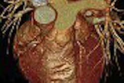
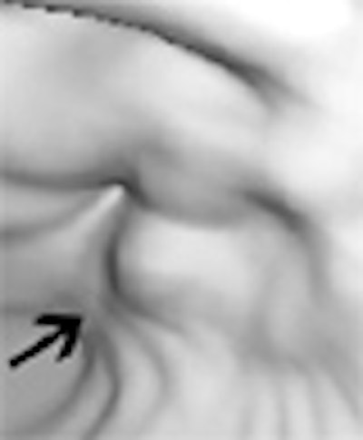
| Early gastric cancer in a 33-year-old woman. There is no identifiable gastric wall thickening on either the transverse CT scan (a, above) or the coronal MPR image (b, below). Virtual endoscopic image (c, bottom) shows a shallow depressed lesion with surrounding mucosal nodularity (arrows) in the angle of the stomach, suggesting early gastric cancer (T1). At histopathologic analysis, this lesion was diagnosed as type IIb + IIc early gastric cancer (pT1). Images used with permission of the Radiological Society of North America (Radiology 2005 Sep;236(3):879-85). |
 |
 |
Both interpretation methods showed a tendency to overstage T1 and T2 cancers, while understaging T3 and T4 cancers. Similarly, it is difficult to evaluate extraserosal invasion of the tumor or its relationship with adjacent organs on CT, the group added, particularly when the interface is nearly parallel to the scanning direction.
"Although MPR images are expected to help overcome this problem, our results with T3 or T4 staging were not satisfactory, and showed a tendency toward underestimation of the tumor stage," the authors wrote.
Overall, for preoperative staging of gastric cancer, MDCT gastrography with multiplanar reconstruction and virtual endoscopy produced better results than transverse CT alone, they concluded.
By Eric Barnes
AuntMinnie.com staff writer
September 26, 2005
Related Reading
Preop chemoradiotherapy beneficial in resectable gastric cancer, July 29, 2004
Study reveals gender, ethnicity differences in esophageal, gastric cardia cancers, May 24, 2004
Extended lymph node dissection does not improve gastric cancer survival, April 4, 2004
Studies compare, optimize contrast in the abdomen, June 28, 2004
Copyright © 2005 AuntMinnie.com