CHICAGO - Location may be more important than size when it comes to managing liver lacerations, according to a study presented Monday at the RSNA meeting.
Bile leaks are a worrisome complication of blunt hepatobiliary trauma, and determining which liver lacerations are serious enough to warrant surgical intervention is difficult work. But it is critical work, inasmuch as bile leaks can seriously affect the morbidity and mortality of trauma patients.
"The ASST (American Society of Surgery and Trauma) guidelines for grading liver trauma following blunt abdominal injury … don't take into account either active bleeding or the presence or absence of bile leaks," said Dr. Brian Lucey, assistant professor of radiology at the Boston University School of Medicine and Boston University Medical Center in Massachusetts. "They really relate to primarily the size of the laceration and hematoma. And it's a very difficult job to try and predict which patients following blunt abdominal trauma and hepatic laceration will actually develop a bile leak."
Most blunt trauma patients undergo CT scans at presentation, he said. If CT patterns could accurately predict which lacerations are likely to result in significant bile leaks, doctors could not only improve their triage, they might also be able to reduce the number of confirmatory cholescintigraphy (HIDA) scans performed, while reserving HIDA scans for patients deemed most likely to have serious leaks.
The group's preliminary results suggest that accurate triage is possible by identifying laceration patterns on CT that are associated with serious bile leaks.
Lucey, along with Dr. Joshua Stuhlfaut, Dr. Jorge Soto, and colleagues, retrospectively examined CT data from 60 blunt or penetrating trauma patients over a one-year period (41 men and 19 women ages 15-83, mean age 36.5 years) with evidence of liver laceration.
CT scans were performed on four-row multidetector-row CT scanner (MX 8000, Philips Medical Systems, Andover, MA) using collimation of 3.2 mm, 50% overlapping reconstruction intervals, following administration of IV contrast material per the facility's standard trauma protocol. No patients received oral contrast. Lacerations were classified by size and location, specifically "central," "peripheral," or "both."
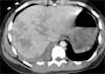
"This (division) is arbitrary, but the central area of the liver we took to represent the hilum, the area where the main bile ducts and blood vessels are coming through, and tried to distinguish that from the peripheral aspects of the liver," Lucey said. "We also decided whether the liver lacerations extended to the medial or lateral edge of the liver, or both. All active bile leaks were documented on HIDA scans.
 |
| The liver was divided into sections for the study. The red line represents the medial surface of the liver. The blue line represents the peripheral surface (P) of the liver. The yellow line roughly separates the peripheral from the central part (C) of the liver. All images courtesy of Dr. Brian Lucey. |
"The reason this is important is that it means you can have a laceration involving the medial and lateral surfaces, but it will remain a peripheral laceration rather than extending to the central portion of the liver," he said.
Results
According to the results, six patients had active bile leaks confirmed on the HIDA scan. Twelve patients had lacerations that included both the medial and lateral surfaces of the liver; the mean size of lacerations that produced active bile leaks was 5.8 cm (with a wide range of 2.5 cm to 10 cm). Five of these 12 patients (42%) had an active bile leak, and the mean size of these lacerations that did not result in an active bile leak was 4.3 cm (again with a wide range from 1 cm to 10 cm).
 |
Twelve patients had lacerations that extended to both the medial and lateral surfaces (mean size 5.8 cm). Five of these patients had an active bile leak (mean size 6.8 cm). Only one patient with a bile leak had an extension to the lateral surface of the liver only.
 |
Eighteen patients had lacerations of the central and peripheral portions of the liver (mean size 6.8 cm), of which 4/18 had an active bile leak (mean size 7.3 cm).
"Lacerations extending to both the medial and lateral surfaces of the liver appear to have the highest incidence of bile leak requiring intervention," Lucey concluded. "And lacerations involving both the peripheral and central parts of the liver seem to have a slightly higher incidence of active bile leak requiring intervention. What we found initially is that location may play a greater role than size in predicting bile leak following hepatobiliary imaging."
Good work, one audience member commented, but bile leakage isn't a concern in many cases, so the question of which cases need intervention isn't entirely resolved by predicting its occurrence. In general, only patients with (ASST) grade 4 and 5 injuries should be operated on, he said.
Session moderator Alec J. Megabow from New York University Medical Center in New York City said the study was an important first step in predicting the need for intervention in liver trauma. He added that future studies would benefit by controlling the timing between the CT exam and the HIDA scan to better assess CT's predictive value.
By Eric Barnes
AuntMinnie.com staff writer
November 29, 2004
Related Reading
Exam technique critical in liver ultrasound, October 22, 2004
Copyright © 2004 AuntMinnie.com


