To a large degree, the initial stability and long-term survival of cementless acetabular cups depends on the bone-prosthesis contact patterns. Unfortunately, orthopedic specialists have yet to find an imaging technique they can depend on to accurately assess these contact patterns.
Radiography is unreliable for evaluating bone-prosthesis support, in-growth, or apposition, wrote Dr. James Howard in a poster presentation at the 2004 American Academy of Orthopedic Surgeons (AAOS) meeting in San Francisco.
More specifically, x-rays are "unreliable for evaluating host bone-implant contact (because of) 2D evaluation. Anterio-posterior and lateral x-ray essentially only provide information on contact throughout a linear section of the cup," explained co-author Dr. John Masonis in an e-mail to AuntMinnie.com. Masonis is with the Miller Orthopaedic Clinic in Charlotte, NC.
"Previous literature has shown that conventional radiographs underestimate gap areas and overestimate the amount of contact of cementless acetabular cups," echoed Howard in an e-mail interview with AuntMinnie.com.
Instead, Howard and his colleagues turned to CT for full cup coverage. "The purpose of this study was to determine the amount and distribution of bone-prosthesis contact for three acetabular component designs using a novel CT image analysis technique," wrote Howard's group, which hails from the London Health Sciences Centre at the University of Western Ontario in Canada.
For this study, the three different types of components were randomly implanted into 18 embalmed, cadaveric hemipelvic specimens. All of the acetabular cups were made of titanium and manufactured by the U.K.'s Smith & Nephew: The Hemisphere has a hemispherical design; the Interfit has a dual-geometry design; and the Trispike has a spiked design.
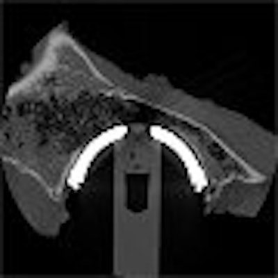
CT imaging was done on a LightSpeed Advantage system (GE Healthcare, Waukesha, WI). A custom, rotational platform was built by the study group in order to manipulate the position of the cup during the scans. The specimens were imaged at 140 kV and 100 mAs at 10° increments.
 |
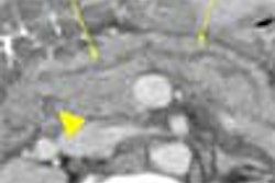
| CT image of an implanted dual-geometry acetabular cup. The majority of the bone-prosthesis contact occurs at the rim of the cup. Image courtesy of Dr. James Howard. |
"For each image, points were mapped on the inner bone surface and the outer prosthetic surface using customized software," the authors said. "The distance from each point on the prosthesis’ surface to the nearest point on the inner bone surface was measured." Contact was defined as bone-prosthesis distance of 0.5 mm or less.
The software (not currently available commercially) was from Enhanced Vision Systems, a Huntington Beach, CA-based firm that manufactures magnification devices for the visually impaired.
According to the results, the total contact measurement (mm2) for the Hemisphere was 2,000 mm2; for the Interfit, 1,000 mm2; and 1,500 mm2 for the Trispike (p < 000.4). The mean percentage of cup contact was 48.6% for Hemisphere, 31.9% for Interfit, and 35.1% for Trispike (p < 0.05).
The authors then used the CT images to create and analyze color maps. They found that with the Hemisphere, the contact pattern was relatively uniform across the cup surface. With the Interfit, CT revealed areas of bone-prosthesis distance near the dome of the cup, but a tighter rim fit. With the Trispike, there was less contact overall.
"The designs analyzed in this study demonstrate the differences in the amount and distribution of bone-prosthesis contact," the group concluded.
While the results were positive, the CT protocol still needs to be refined, Masonis said.
"We utilized this technique only for the purposes of this study and additional stem contact studies. We do not employ this technique clinically on patients. The elevated radiation exposure could also be a potential issue for live subjects. Therefore it was only performed in cadaver specimens," he said.
Future studies will focus on the radiation dose to the patient, as well as cup orientation and the effects of artifacts from the ipsilateral femoral head/stem, Howard said.
In related research, a two-part article in the June 2004 issue of the American Journal of Roentgenology will expound on acetabular imaging. Dr. John Harris, Jr. and colleagues from the University of Texas-Houston Medical School will discuss acetabular fractures, including a new definition of the letournel anterior column, as well as a CT-based reclassification of these types of fractures.
By Shalmali PalAuntMinnie.com staff writer
April 28, 2004
Related Reading
Imaging documents damage associated with NSAIDs usage, November 27, 2003
Proper positioning for the pelvis and proximal femur, August 8, 2003
Copyright © 2004 AuntMinnie.com