VIENNA - CT and MRI, both anatomical and functional, can be combined to improve the targeting of radiation therapy of the prostate, according to researchers from the University of Nijmegen in the Netherlands. At today’s prostate imaging sessions of the European Congress of Radiology, Jorn van Dalen, Ph.D. presented a study utilizing the novel coregistration method the group created.
"MRI gives you an accurate visualization of the anatomy, but it also provides important functional information which improves tumor localization -- you can think of dynamic or fast MRI and also MR spectroscopy," van Dalen said. "However, we also need CT for radiotherapy treatment planning, to calculate the dose distribution. In order to incorporate this MRI into treatment planning, we have to fuse it accurately with CT, particularly (in) intensity-modulated radiation therapy (IMRT), where you can give a decreased dose in the surrounding tissue and an increased dose on the boost target."
 |
| Dynamic (left) and functional (center) MRI of the prostate can improve tumor localization. All images courtesy of Jorn van Dalen, Ph.D. |
IMRT accuracy is generally within 2 mm in order to minimize the side effects associated with the irradiation of healthy tissue, he said, so 2 mm was also the benchmark used to evaluate the feasibility of the fusion method used by investigators from the university’s radiology, physics, and urology departments.
In the study, 20 patients with histologically proven prostate cancer underwent both multislice CT acquired with the use of an endorectal balloon and 50-minute MRI examination using a 1.5-tesla MRI scanner with pelvic phase array and rectal coils. The MRI exams included:
- T2*-weighted MEDIC protocol for optimal marker visualization
- T2-weighted MRI in three planes for optimal anatomical visualization (0.6 x 0.6mm)
- Dynamic Gd-DTPA-enhanced MRI, 7 slices/2 sec
- 3-D 1H-MR spectroscopy imaging
"Gold markers (1 x 7 mm) were inserted into the prostate to ensure the best fiducials for the fusion process," he said. "The automatically segmented parts are then fused using a so-called ICT method, the Intake Closed-Point method."
 |
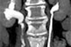
| Gold markers (center) before (left) and after (right) ICP coregistration method is applied in 2-D images. |
MRI and CT were performed within a week of each other. The results were validated by measuring interobserver and intraobserver variability, and the fiducial registration error, which is the distance between the markers after the fusion process. The most important measure, the target registration error, is the difference between homologous points developed by Fitzpatrick et al in 1998, rather than the distance between the markers, van Dalen said.
 |
| Anatomic MRI with CT (left) and functional MRI with CT (right) show tumors and markers. |
In all, four operators performed 80 fusions. Interoperator variability at the center of the prostate was 0.3 mm, 0.7 mm at the rim, with statistical outliers demonstrating greater than 3-mm variability at the rim occurring in 4% of the fusions, he said. This gauge was significantly larger than the fiducial localization error: 0.3 mm for CT and 0.9 mm for MRI.
"The last (measure) is the target registration error: 1.2 mm at the center of the prostate and increasing to 2.4 mm at the rim of the prostate," van Dalen said. "Most of it (>82%), is due to non-rigidities between the two images.... Smaller (error) contributions are coming from MRI distortion, marker migration, and fiducial localization," he said.
 |
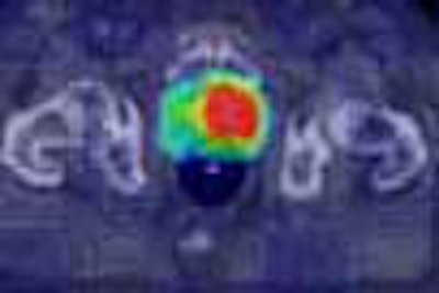
| Clear indications of tumor are seen in anatomic (left) and functional (center) MRI. When fused with CT image (right) for IMRT, higher radiation doses can be more accurately targeted to the tumor, sparing surrounding tissues. |
The gold markers and the fusion method enable accurate fusion of high-resolution anatomic and functional MR and CT to within about 2 mm, van Dalen concluded, allowing the addition of anatomical and functional MRI data to IMRT treatment planning.
AuntMinnie.com staff writer
March 5, 2004
Related Reading
Screening for prostate cancer leads to earlier detection, January 28, 2004
Age bias may influence treatment of localized prostate carcinoma, January 22, 2004
Hormone therapy for prostate cancer may cause rapid bone loss, January 19, 2004
Copyright © 2004 AuntMinnie.com