CHICAGO - For standard head CT applications such as initial screening, standard exposure protocols help ensure the best possible image quality when it's needed. But for non-critical applications such as routine follow-up, minimizing patients' radiation exposure makes more sense. In today's dose-conscious environment, researchers are examining the trade-off between dose and image quality, and finding that lower-dose protocols don't necessarily reduce the diagnostic utility of the data.
As part of an institution-wide study that halved the radiation dose in many common CT applications, neuroradiologist Dr. Peter Bove and his former colleagues from Massachusetts General Hospital in Boston evaluated 20 non-CNS cancer patients, all over age 65, with both standard and half-dose CT of the head.
The goal was to compare the both the quality and potential clinical applicability of low-mA head CT exams with that of conventional exams.
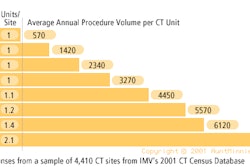
"At our institution we routinely get one or two head CTs a day; many of the patients have had angiography or other procedures, so reducing the mA in those patients is really critical," Bove said in a presentation Monday at the neuroradiology sessions of the RSNA meeting.
All of the patients underwent conventional non-contrast head CT using a standard multidetector-row technique (LightSpeed premium multislice scanner, GE Medical Systems, Waukesha, WI). The protocol (170 mA, 140 kV, pitch 3, table speed 7.5 mm/sec.) yielded a radiation dose of approximately 65 mGy (6.5 rad).
Having offered their informed consent, the patients then underwent a second scan comprising four additional sections. The protocol was identical to that of the first scan except for the mA, which was nearly halved to 90. Dose being proportional to mA, the effective dose was also nearly halved, Bove said.
The researchers evaluated representative attenuation ROIs in identical gray- and white-matter locations for both sets of images using a five-point scale, 5 being better than 170 mA and 1 being non-diagnostic. For each ROI, they compared image noise, gray and white matter conspicuity, subarachnoid sharpness, ventricular margins and posterior fossa in both sets of images.
The results were also assessed quantitatively. The image contrast-to-noise ratio was defined as [mean GM ROI- mean WM ROI] / [( standard deviation GM ROI)2 + (standard deviation WM ROI)2]1/2. Gray vs. white matter conspicuity was defined as (mean GM ROI- mean WM ROI) / (mean WM ROI). The data were analyzed using the student's T-test for unequal variances.
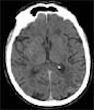
In the visual analysis, the low-dose images showed "an increase in modeling, and we can also see that the sharpness is not quite as good. You can see that the margins aren't quite as sharp," Bove said.
Yet most structures remained well defined, and no images were non-diagnostic, he said. The gray matter was not significantly less conspicuous in the half-dose (0.41 ± 0.03) vs. the normal-dose (0.39 ± 0.19) images (p=0.32).
 |
quality remains acceptable for most applications. Image courtesy of Dr. Peter Bove.
As for the signal-to-noise ratio, noise increased by approximately 22% in the half-dose images (1.39 ± 0.38) compared to the normal-dose images (0.39 ± 0.19) (p=0.32). Subjectively, the overall image quality "is only considered slightly less ... than 170 mA, which is the standard," Bove said.
"The 90 mA scans probably have anatomic resolution sufficient for certain routine evaluations, and that is, to follow gross morphological changes and complications, follow-up of hydrocephalus, especially in kids.... This is also to say that the technique is probably not good for all cases, the anatomic resolution not good enough for examining the brain as initial screening, or when you're following up small subtle lesions" such as lacunar infarct, early cortical stroke, or petechial hemorrhage, he said.
The moderators and the audience had plenty of ideas for follow-up studies. One observer wondered how well the low-dose images would hold up in the watery myelin and overall different water environment of children's brains. Another asked if creative windowing and postprocessing techniques might improve visualization of the low-dose images even further. GE is working with the researchers at MGH to develop new visualization techniques, Bove said.
By Eric Barnes
AuntMinnie.com staff writer
November 27, 2001
Copyright © 2001 AuntMinnie.com