Radiation exposure can be lowered from 280 mAs to 120 mAs without affecting diagnostic image quality in unenhanced chest CT scans, according to findings presented in the latest issue of the American Journal of Roentgenology.
Dr. James G. Ravenel and colleagues from the SUNY Upstate Medical University of Syracuse in New York obtained 10 sets of images from eight patients who underwent CT-guided chest biopsies. Imaging was performed at 120 kVp on a CT/i scanner (GE Medical Systems, Waukesha, WI).
According to the study, "scans were obtained using 5-mm collimation and a pitch of 1.5:1 to generate a set of four to five helical images using a 5-mm reconstruction interval and a 'detail' reconstruction algorithm" (AJR, August 2001, Vol. 177, pp. 279-284).
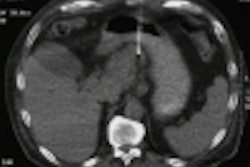
Researchers acquired, from each patient, six images of the same region exposed at doses of 40, 80, 120, 160, 220, and 280 mAs. CT data was used to reconstruct sections with a reduced field of view, including the hemithorax contralateral to the biopsy site, the mediastinum, and a small portion of the ipsilateral hemithorax.
Five radiologists evaluated the scans, and ranked them according to image quality. Observers were explicitly advised to base image quality on mottle, which "depends on the total number of x-ray photons used to generate the CT," the study said.
In interpreting the scans, the radiologists were unable to differentiate image quality generated at 160 mAs from those at 220 and 280 mAs. On the other hand, they were able to perceive differences in quality at 40, 80, and 120 mAs.
"[A chest CT image produced at 160 mAs] was identified as the borderline technique value. At a technique factor equal or greater than 160 mAs, image quality is similar; at less than 160 mAs, image quality is progressively identifiable as inferior," the study said.
Images generated at 160 mAs were used as references by radiologists to evaluate quality at the five remaining exposures.
When compared with the reference image, the difference in image quality at 120 mAs was negligible. Radiologists did not consider the difference significant enough for additional radiation. At 80 mAs, a slight reduction was seen, but observers deemed the images sufficient and did not recommend more exposure. At 40 mAs, however, image quality was considered inferior to the reference image, leading radiologists to suggest higher radiation at this technique factor.
"A reduction of the tube current to 120 mAs for routine unenhanced chest CT examinations would appear justified and would reduce the typical adult effective dose from 6.0-2.6 mSv. This decrease corresponds to a 57% reduction in patient dose with no adverse impact on image quality," the study stated.
Based on the findings, CT doses can and should be lowered, Ravenel said.
"Radiologists and imaging professionals who are responsible for developing scanning protocols need to address the tradeoff between patient dose and image quality for each diagnostic imaging study," Ravenel wrote. "The balancing of dose and image quality should be performed explicitly to ensure that patient doses are kept as low as reasonably achievable."
Others have reached similar conclusions concerning reduced radiation levels and their effect on diagnostic image quality.
In an accompanying commentary, Drs. Edward Nickoloff and Philip Alderson offer several techniques for lowering CT exposure while providing quality scans. The authors suggest reducing amperage and scan time, using thicker CT slices, and spacing CT slices at pitches greater than 1:0 in single-slice axial or helical CTs (American Journal of Roentgenology, August 2001, Vol. 177, pp. 285-287).
Dr. John Haaga agreed in a second commentary, adding that CT exposure can be reduced by increasing the pitch in helical scanning from 1.0 to 1.5. In addition, he recommends adjusting the milliampere-second requirement to the diameter of the patient rather than to the weight, because "the diameter better correlates with the distance of the pathway traversed by the x-ray beam" (American Journal of Roentgenology, August 2001, Vol. 177, pp. 289-291).
Haaga said the techniques reduce radiation exposure by 30%-50%.
By Jennifer LungAuntMinnie.com staff writer
August 7, 2001
Related Reading
Slashing CT dose in clinical practice, July 25, 2001
CT over the rainbow: color-coding protocol reduces errors, May 30, 2001
FDA's radiation concerns may lead to dose displays for scanners, May 22, 2001
Study sparks fears over kids’ exposure to CT radiation, February 13, 2001
CT radiation exposure higher than necessary in children, January 23, 2001
Copyright © 2001 AuntMinnie.com