CT has come a long way since 1972, when Godfrey Hounsfield built the first scanner in his lab at EMI. The next few years saw improved resolution, faster acquisition, and machines that could cover more anatomic territory with fewer motion artifacts, trends that have continued with each successive generation of scanners.
The introduction of spiral CT scanning with volumetric imaging in 1991 heralded a revolution in diagnostics, with better visualization of small pathologies, and for the first time, data reconstruction techniques tailored to specific diagnostic needs. Faster gantry rotation, more powerful x-ray tubes, and better interpolation algorithms have sharpened the picture even more since then.
But no innovation has advanced the modality more than the 1998 introduction of multidetector-row CT (MDCT) -- and no diagnostic procedure will benefit from it more than CT angiography, according to Dr. Geoffrey Rubin, professor of radiology at Stanford University School of Medicine in Stanford, CA.
At the June International Symposium on Multidetector-row CT in San Francisco, Rubin waxed enthusiastic about MDCT's benefits in angiography, noting its superior speed, anatomic coverage, and spatial resolution compared with single detector-row scanners (SDCT) and the current gold standard, digital subtraction angiography (DSA).
In a study of 48 patients published in the June issue of Radiology (Radiology 2000;215:670-676), Rubin and colleagues found that for coverage areas of similar length, MDCT reduced overall scan time by 60% (from 75 sec. to 30 sec.) while enabling 40% thinner collimation.
"What is particularly striking is that the contrast-medium dosage dropped from an average 320 ml down to 115 ml, a reduction of 64%. And even though we've reduced our contrast dose...the average attenuation within the aorta and iliac arteries did not change statistically significantly," he said.
While scan time was 2.5 times faster with MDCT than with SDCT, scan efficiency (speed divided by section thickness) was adopted as a more relevant measure than speed because it accounts for varying slice thickness between scans. Overall scan efficiency was 4 to 4.1 times greater with MDCT, "which makes a lot of intuitive sense given the fact that we have four detector rows rather than one," Rubin said.
The team also found that contrast efficiency, defined as average aortic attenuation divided by the number of grams of iodine used, was 2.4 times greater with MDCT than with SDCT.
"This is a very important result considering the relative risk of the delivery of iodinated contrast to patients with vascular disease," he said.
Rubin showed chest images acquired with SDCT that had significant pulsation artifacts in the heart, the left pulmonary artery, and the descending aorta. Artifact was greatly reduced in the MDCT images, which also delivered better branch visualization, better delineation and detection of calcified plaque, and greater homogeneity of vascular and parenchymal opacification, Rubin said.
An artifact that does remain in MDCT-acquired chest images can actually be useful in diagnosing ischemia, he said. A result of displacement of the intimal flap over the 20-30-second exposure time, the stairstep artifact visible in such images corresponds to the systolic and diastolic phases of heart, he said.
"It's a well known phenomenon ... that when the pressure of the true lumen goes down, you have expansion of the false lumen, which can compress the true lumen during systole and limit blood flow down into the abdomen -- the so-called inflow limitation. Patients can present with ischemia of the visceral organs simply on the basis of expansion of the false lumen in diastole."
The phenomenon can't be seen in MRA with gadolinium contrast, he said, as the intimal flap would be visualized as an average value over the course of the scan.
In MDCTA of a diabetic patient with a high creatinine level, Rubin took the unusual step of using gadolinium contrast, a paramagnetic agent with no contraindications except for a history of prior reaction.
"Because I can complete the CT scan in 12 seconds, I can load the power injector with 40 ml of gadolinium...and get diagnostic-quality CTA without using any iodine whatsoever," he said.
The group is now conducting a prospective trial to assess the utility of CT angiography with gadolinium in patients who cannot undergo MRA.
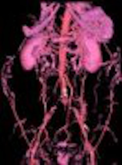
MDCT has also spurred development of several new applications of CTA, such as evaluation of lower extremity occlusive disease. MDCT can cover a distance of 130 cm in less than 70 seconds, enabling complete acquisition of inflow and run-off vessels, Rubin said.
"One of the challenges of imaging severe lower extremity lower occlusive disease with the gold standard, DSA, is that when there is asymmetric disease, it can be very difficult to achieve simultaneous opacification of both legs to be able to make a complete assessment."
This isn't a problem with MDCT, which can visualize branch patency over greater distances, he said.
"The localized injection of the DSA frequently results in blood shunting around obstructions and going along the path of least resistance. Simultaneous opacification of the entire blood pool with the peripheral venous injection in MDCT allows better visualization distal to occlusions," Rubin said.
In 16 patients with lower extremity claudication or limb-threatening ischemia evaluated so far, MDCTA correctly identified all 52 occluded segments and patent segments with stenoses greater than 50%, he said.
MDCT's ability to scan longer segments is also valuable in planning endoluminal repair, when knowing where the aneurysm begins is not as important as knowing where the outside wall of the vessel is.
In heavily calcified vessels, planar reformations of the CT data based on the flow lumen can characterize the residual lumen effectively. With appropriate window-level settings -- an essential component, Rubin said -- calcification is also well visualized with MDCT.
Finally, MDCT's superior volumetric spatial resolution enables assessment of complex lesions that are not adequately visualized DSA or single-detector CTA, he said. MDCT's ability to map out the entire network of vessels with volume rendering and 3-D views is a valuable planning tool for interventional radiologists about to embark on long embolotherapy sessions, Rubin said.
"(MDCT) can perform scans faster to get more anatomic coverage with less contrast, and we're able in some cases to even eliminate the use of iodinated contrast. We're able to study vascular beds that we could never even see in their entirety before, such as lower-extremity runoff, and we're evaluating vascular beds...with spatial resolution that is simply unprecedented by prior methods," Rubin said.
By Eric BarnesAuntMinnie.com staff writer
August 4, 2000
Let AuntMinnie.com know what you think about this story.
Copyright © 2000 AuntMinnie.com