Researchers at Northwestern University are developing a new tool for cardiovascular disease with their work on echogenic immunoliposomes (ELIPs) -- injectable microspheres that go beyond current ultrasound contrast agents by surface binding with their targets.
Developed by Dr. David McPherson, a professor of cardiology and director of Northwestern’s Echocardiography Laboratory in Chicago, ELIP imaging could enhance the general knowledge of atherosclerotic plaque development. The liposomes could also enable the targeting of drug or gene therapy toward specific atherosclerotic sites without damaging cells.
"While our research is in the early stages, we believe that our combined technology will have far-reaching implications in humans, allowing for more directed atherosclerotic and thrombolytic therapy," McPherson said.
Upon reaching their arterial target, ELIPs produce an acoustic shadow, enhancing ultrasound’s capacity to visualize plaque in the artery. The Northwestern group’s technique involves the conjugation of various antibodies to the surface of the liposomes, "which will then attach to the target molecules in atherosclerotic blood vessels," said co-investigator Ashwin Nagaraj, Ph.D.
"Since different molecules are expressed at different stages of the disease in endothelial cells, this also provides the ability to diagnose the extent of atherosclerosis," noted Nagaraj, a research engineer at the Northwestern lab.
In an earlier publication, the Northwestern researchers described the systemic ultrasound enhancement of a left ventricular thrombus after the intravenous injection of ELIPs conjugated to anti-fibrinogen (Circulation, June 11, 2002, Vol.105:23, pp. 2772-2778).
 |
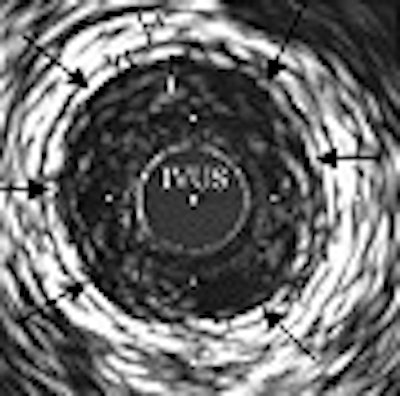
| Intravascular ultrasound (IVUS) images of a porcine right femoral artery showing enhancement of injured endothelium by anti-fibrinogen-conjugated echogenic immunoliposomes (ELIPs). Images were acquired after saline injection (a) and five minutes after injection of an 8 mg dose of anti-fibrinogen-conjugated ELIPs (b). Arrows indicate enhanced endothelium. I = intima; M = media; A = adventitia. Image courtesy of Ashwin Nagaraj, Ph.D. |
The ELIPs themselves are composed of four primary lipids -- PC, MPB-PE, PG, and cholesterol -- and are made by a lyophilization process, according to the Circulation article. By manipulating the component lipids, the Chicago lab had increased the echogenicity of ELIPs by around 300%.
ELIPs can also be prepared to carry drugs, such as antibiotics, thrombolytic drugs or gene therapy, which, in combination with ultrasonic pulses, are released at the site of a plaque or a clot or into living cells. This cavitation process, with ultrasound increasing the energy of the microbubble, then opens the cell membrane, allowing drugs to enter.
The investigators are working on a way to trap the appropriate drugs or genes in the conjugated liposomes for release at the disease site with low-frequency ultrasound, Nagaraj said.
By Bruce SylvesterAuntMinnie.com contributing writer
March 1, 2004
Related Reading
ISET talks offer innovative ways to survey aortic aneurysm repair, January 3, 2004
Affordable equipment accelerates ultrasound proliferation, January 26, 2004
External therapeutic US accelerates thrombolysis in heart attack patients, December 9, 2003
Contrast-enhanced ultrasound not just for cardiac applications, May 14, 2003
Copyright © 2004 AuntMinnie.com