Ever since Wilhelm Conrad Roentgen's discovery of x-rays in 1895, the field of medical imaging has continued to expand across a multiplicity of applications, and one of the most fascinating is its use as a forensic tool to determine the causes of trauma, both in living and deceased individuals.
Forensic radiologists and radiologic technologists apply their expertise to imaging exams -- typically x-ray and CT -- intended to illuminate signs of trauma. So how do these experts approach forensic radiology? AuntMinnie.com spoke to two experts about the state of the specialty.
Training is key
What is forensic radiology? Put simply, it's the practice of using imaging for medicolegal purposes, according to Jamie Elifritz, MD, chief medical officer of the Forensic Radiology Group, a national company focusing on service work and forensic imaging education. Postmortem imaging involves deceased individuals and may be applied to investigations of cause of death, she said.
"Imaging technology has become more affordable and attainable, and this allows medical examiner’s offices to incorporate advanced imaging in their practices," Elifritz told AuntMinnie.com. "There's also increased interest among forensic pathologists about incorporating forensic imaging in their investigations."
Elifritz was mentored by the "father of forensic radiology" Gil Brogdon, MD, during her residency at the University of South Alabama in Mobile, AL, and trained under forensic radiologist Gary Hatch, MD, during her fellowship at the University of New Mexico/Office of the Medical Investigator (OMI) in Albuquerque. In 2020, she was tapped to serve as the director of forensic imaging at the OMI; while there, she interpreted cases for the clinical service, developed an educational program for the forensic pathology fellowship, and participated in research. She also serves as chair of the guidelines committee for the International Society of Forensic Imaging (ISFRI).
 Jamie Elifritz, MD, chief medical officer of the Forensic Radiology Group.
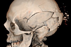
Just as pediatric imaging isn't simply imaging of "small adults," forensic imaging is its own subspecialty, according to Elifritz. She outlined the complexity of forensic reporting, citing a gunshot wound as an example: Entry wounds in the skull "typically show internal beveling, but there are nuances -- specifically with keyhole types of entry -- and this determination should be made in conjunction with the external exam. There are significant legal implications," she said.
"The task of reading forensic imaging is complex," Elifritz noted. "There is a learning curve for radiologists to understand postmortem artifacts."
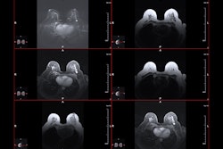
Feedback from a forensic radiologist like Elifritz can change how a medical examiner proceeds with a case. Below is an image of an individual who, given his nonsuspicious history, toxicology results, and external examination findings, would typically not have undergone autopsy. Elifritz noted that the mild degree of decomposition was inconsistent with the degree of bowel dilation. She followed the bowel on CT and identified an inguinal hernia-containing bowel with evidence of obstruction. Given these findings, the decedent underwent autopsy, where a strangulated hernia was identified.
 The axial images on soft tissue windows demonstrate the dilated bowel, a common finding in decomposition. The images with the circles denote the incarcerated hernia. The larger photo is the incarcerated and strangulated bowel at autopsy. Images and caption courtesy of Jamie Elifritz, MD.
"The degree of bowel dilation was greater than expected for mild decomposition and when I followed the bowel, I found the hernia-containing bowel – which is a common radiological diagnosis but in this case [illuminated the cause of death]," she told AuntMinnie.com.
The most important factor in forensic imaging is the partnership between forensic pathologists and forensic radiologists, and if radiologists want to be of service to forensic pathology colleagues, they must understand autopsy and medicolegal investigations, according Elifritz.
"I would encourage radiologists who are interested in forensic/postmortem imaging to look into ISFRI, NAME [the National Association of Medical Examiners], and the AAFS [American Academy of Forensic Sciences] for training and education opportunities," she said.
State of the art
Where is the specialty today? It has grown, but there's more to be done in terms of training, radiologic technologist and doctoral candidate Kristin Beinschroth of the Gurnick Academy of Medical Arts in Van Nuys, CA, told AuntMinnie.com. Beinschroth is the program director of the x-ray technician with medical assistant skills program at Gurnick. In a presentation she delivered March 9 to the California State University Northridge Bachelor of Radiologic Sciences Alumni Association, Beinschroth detailed the current state of forensic imaging.
"Forensic radiology is a subspecialty dedicated to antemortem and postmortem imaging, usually for medicolegal purposes, and is increasingly used in conjunction with conventional autopsy as a virtual autopsy, which connects radiology with forensic medicine," she said.
 Kristin Beinschroth of Gurnick Academy of Medical Arts.
The specialty is used in anthropology to date and assess mummified remains; in criminology to determine the nature and causes of crime, whether trauma was accidental or not and to identify the point of impact of a weapon and its shape; and to classify skeletal remains and perform facial reconstruction. Technologists perform the exams and radiologists interpret the results, then work with a multidisciplinary team that may include laboratory technicians, pathologists, coroners, and lawyers, she said.
Beinschroth described a case in which a three-day-old infant presented with bruising over the clavicle, a finding that could indicate "nonaccidental trauma." The baby underwent x-ray imaging, which showed skull fractures, and an investigation began. But then the mother revealed that she had been in a car accident on the day her child was born. She had a full-body trauma CT scan that confirmed her report.
"Investigators found injuries in the mother on CT that indicated that the child's injuries were due to the accident, not abuse," Beinschroth said.
Researchers are actively exploring how best to use forensic radiology. In a study published March 18 in Forensic Science International, a team led by Katharine Kolpan, PhD, of the University of Idaho in Moscow explored the use of CT-based augmented reality (AR) for reconstructing fractured, fragmented, and damaged craniofacial remains in forensic anthropology, finding that "CT-based AR craniofacial reconstruction can reduce bone damage from overhandling," and that "AR allows the viewer to assess the skeleton in three rather than two dimensions."
"[Using CT with AV] allows forensic anthropologists a means of assessing aspects of the biological profile and traumatic injuries without risking further damage to the skeleton," the group explained. "Additionally, the holographic images can be used to explain complicated concepts in a courtroom without the emotional response related to using bony elements as courtroom exhibits."
One of the most exciting uses of imaging for forensic purposes is the virtual autopsy, according to Beinschroth.
"Virtual autopsy is noninvasive, doesn't harm the body, doesn't tamper with forensic evidence, and can be used in cultures and situations where conventional autopsy is taboo," she noted.
Both Elifritz and Beinschroth have developed educational resources for forensic imaging and hope to continue these efforts.
"I plan to be involved in developing more education for forensic pathologists," Elifritz said. "There may be opportunities for dual fellowships in forensic imaging -- a dual pathway for radiologists and pathologists."