The number of artificial intelligence (AI) algorithms cleared for use in neuroradiology and musculoskeletal radiology has surged recently, and "judicious" adoption of these tools will likely improve quality and efficiency, according to a new analysis.
After reviewing peer-reviewed studies and the current range of AI products cleared by the U.S. Food and Drug Administration (FDA) for these applications, researchers led by Dr. Elisa Berson of Yale School of Medicine noted a recent surge in clearance of AI clinical tools to accelerate and enhance diagnosis and treatment since 2018, covering a broad spectrum of areas.
"While many approved products are applied for automated quantification, there are increasing numbers of approved products that move beyond quantification to detection and even diagnosis," the authors wrote in their article published in Seminars in Roentgenology. "As FDA approvals of AI technology continue to increase, it will be increasingly important for radiologists to be aware of the product landscape and play a role in the selection and application of these products in the reading room."
As of August 2022, when the study was conducted, the FDA had issued 510(k) clearance for almost 200 algorithms -- many pertaining to neuroradiology and musculoskeletal radiology -- and many are being integrated gradually into clinical practice. The researchers listed the software, product functions, modalities, and clearance dates, as well as peer-reviewed literature evaluations where available.
Image quantification is among the most common applications for AI, providing automated segmentation and measurement of brain volumes, for example.
Quantification
In their paper, the researchers described Belgian company Icometrix's portfolio of AI products that quantify the area and/or volume of brain regions on CT or MRI to inform clinical care and track disease processes over time. They also cited evidence that these volumetric measurements have "proven correlations with multiple sclerosis, dementia, and epilepsy which can guide radiologists, neurologists and neurosurgeons."
Other products that aim to automatically quantify brain regions and pathologic changes (such as white-matter hyperintensity) include NeuroQuant (Cortechs.ai) and Quantib ND (Quantib).
The authors say that the "shared value proposition" of these types of AI products is in providing quick quantification and comparison of patient-specific brain region measurements to those of reference populations or prior scans from the same patient and presenting the information so that it is easily understood by clinicians and patients.
Such quantitative AI products are "easily integrated with clinical workflow and add valuable information through automated segmentation and measurements to inform clinical practice and patient care," the researchers wrote.
Image optimization
Berson and colleagues also discussed examples of AI applications in image optimization, such as AlgoMedica's PixelShine technology for denoising of low-dose CT scans. They noted that AI has the potential to enhance details and clarity of lower-resolution and lower-radiation scans without losing diagnostic utility.
In addition, they highlighted research on patients with lung nodules in which CT images that were reconstructed using deep-learning and model-based iterative reconstruction "significantly improved" subjective scoring on image quality when compared with other low-dose images.
Improved visualization, detection
The authors also described how a series of AI-based software "improve visualization" of volumetric scans via 3D reconstruction or generation of synthetic CT images from MRI scans.
Furthermore, AI programs can be designed to intake information and perform segmentations and measurements to help in detection and diagnosis, according to the researchers.
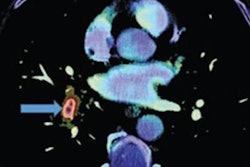
Although the majority of software for detection of intracerebral hemorrhage, large vessel occlusion (LVO) stroke, and aneurysms are primarily cleared for triage purposes by the FDA, some AI products are specifically cleared for diagnostic applications. These include Nicolab's StrokeViewer software -- the first product to be cleared by the FDA for the AI diagnosis of acute ischemic infarct, according to the researchers.
AI in MSK radiology
Although not as many algorithms have been cleared for musculoskeletal (MSK) applications as there have been for neuroradiology, the software that's currently available is designed to handle tasks such as fracture detection, diagnosis and quantification of knee arthritis, and quantification of spinal degenerative disease.
Missed fractures on plain radiographs are a common diagnostic error. The review looked at how AI products, such as BoneView (Gleamer) and FractureDetect and OsteoDetect (both from Imagen), are being used to assist in fracture detection from plain radiographs.
For BoneView, a study by Guermazi et al examined 480 patients with a total of 350 fractures (30 x-rays with and without fractures for 8 body regions) and reported 64.8% sensitivity without AI assistance and 75.2% sensitivity with AI assistance and a corresponding specificity of 90.6% without AI and 95.6% with AI, respectively. These accuracy improvements were achieved with a significantly shorter reading time by over six seconds.
Osteoarthritis
The authors also shared their analysis of the evidence and supporting data for AI products that aim to streamline scoring and reporting of common chronic diseases such as osteoarthritis based on standing anteroposterior (AP) radiographs of the knee, such as the Knee Osteoarthritis Labeling Assistant (KOALA) by Image-Biopsy Lab.
In a study comparing KOALA-assisted vs unassisted reading of knee osteoarthritis, for all three categories (sclerosis, osteophyte, joint space narrowing), interreviewer agreement between physicians in detection and Osteoarthritis Research Society International Scores increased after using KOALA, according to the researchers.
The researchers noted that their review did not include an evaluation of the compatibility of various imaging systems with the AI software. Although most vendors try to have their software be as broadly applicable as possible, some AI products are only available for certain imaging technology vendors, according to the authors.
Assessment, monitoring
On the downside, the researchers observed that plans for continued assessment or mechanisms for feedback reporting by radiologists are "rarely described."
They said that AI products should be continuously assessed for their accuracy and applicability to different patient populations and care settings, to ensure that practicing radiologists can report "quality gaps" and allow technical adjustments to take place where necessary.
"It is important to not only be aware of the variety of tools available but also to evaluate the original validation data and compare their results with studies reporting real-time use of AI in populations and care settings, which may differ from the development data," they wrote.
The authors concluded that "AI products should undergo continued assessment of their accuracy and applicability through continued learning, validation, and adjustment as the technology is adopted across different institutions and countries and applied to different patient populations. In the future, more systematic evaluation and mechanisms for monitoring of AI algorithms and reporting algorithm drift or other quality gaps would be desirable."
Ultimately, though, "it is likely that judicious adoption of AI will enable radiologists to provide higher quality and more efficient care for their patients," the researchers wrote.