Chinese researchers have found that artificial intelligence (AI) outperforms not only radiologists but also clinical and radiomic models when it comes to predicting lymph node metastasis in patients with pancreatic ductal adenocarcinoma cancer.
The results show promise for making a particularly challenging clinical task easier, wrote a group led by Dr. Yun Bian, PhD, of Changhai Hospital in Shanghai. The research was published on September 6 in Radiology.
"Preoperative identification of lymph node metastasis still mainly depends on the radiologist's report," it noted. "However, the lower [area under the receiver operating characteristic curve] values ... reported in [our] study show that lymph node metastasis prediction by radiologists remains extremely difficult."
Assessing the lymph nodes of patients with pancreatic ductal carcinoma is critical for setting an accurate prognosis and guiding patient care, the team explained. But predicting lymph node metastasis before surgical treatment of pancreatic cancer is tricky: Studies have suggested that CT only has a sensitivity of 25% and positive predictive value of 28%. And although radiomics have shown promise, its efficacy is unclear -- especially since radiomics features for pancreatic cancer have been taken from tumors rather than lymph nodes.
Bian's team sought to explore whether an AI algorithm could improve CT's performance for presurgical prediction of lymph node metastasis in patients with pancreatic cancer via a study that included 734 patients diagnosed with pancreatic ductal adenocarcinoma who underwent CT imaging between January 2015 and April 2020. The group developed and validated a preoperative AI algorithm that used CT data to predict metastasis in individuals with the disease.
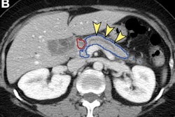
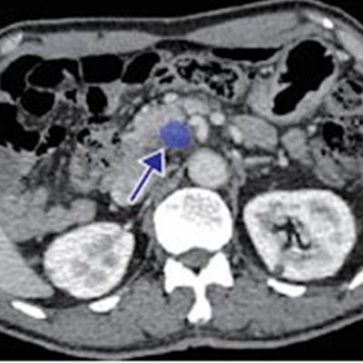
 Images in a 71-year-old man with pancreatic ductal adenocarcinoma in the lymph node-negative group. (A) Hematoxylin-eosin stain shows that the peritumoral lymph nodes (arrows) are negative (hematoxylin-eosin staining; magnification, 31). The dashed line indicates the tumor periphery. (B) Graphic shows lymph nodes (green arrows), tumor (blue arrow), and pancreas (yellow) segmented and diagnosed by artificial intelligence (AI). (C) Axial portal-phase CT scan segmented by the AI model shows a negative lymph node (arrow) located at the pancreatic head. (D) Axial portal-phase CT scan segmented by the AI model shows a negative lymph node (arrow) located at the pancreatic head. (E) Axial portal-phase CT scan segmented by the AI model shows an infiltrative low-attenuation mass (arrow) at the pancreatic head.
Of the study cohort, 545 cases were used to train the algorithm and 189 to validate it. The researchers then used the area under the receiver operating characteristic (AUC) curve to compare the performance of the AI model with a clinical model and a radiomics model (both of which included information such as tumor size and differentiation grade).
The artificial intelligence algorithm outperformed not only radiologist readers for predicting lymph node metastasis in patients with pancreatic cancer across a variety of measures -- radiologists showed an AUC of 0.65 on the validation set compared to the algorithm's validation set AUC of 0.92 -- it also bested the clinical and radiomics models.
| Performance by AI model for predicting lymph node metastasis in patients with pancreatic cancer (AUC) | |||
| Prediction of lymph node metastasis | Clinical model | Radiomics model | AI model |
| Training set | |||
| AUC | 0.76 | 0.71 | 0.91 |
| Accuracy | 71.4% | 67% | 85% |
| Negative predictive value | 70.8% | 72.1% | 85.2% |
| Positive predictive value | 72.3% | 62.6% | 84.7% |
| Sensitivity | 62.6% | 72.4% | 82.7% |
| Specificity | 79% | 62.2% | 89.3% |
| Validation set | |||
| AUC | 0.77 | 0.68 | 0.92 |
| Accuracy | 73% | 63% | 85.2% |
| Negative predictive value | 79.6% | 73.9% | 84.4% |
| Positive predictive value | 67.3% | 56.7% | 86.3% |
| Sensitivity | 79.1% | 79.1% | 80.2% |
| Specificity | 68% | 49.5% | 89.3% |
Bian and colleagues' work could improve pancreatic patient care, wrote Dr. Linda Cho and Dr. Elliot Fishman, both from Johns Hopkins University in Baltimore, in a commentary published with the study.
"AI has the potential to improve staging and our understanding of tumor biology, and to help select the patients most likely to benefit from surgery and tailor the most appropriate neoadjuvant or adjuvant therapy," they noted. "In conjunction with current efforts focused on earlier detection of pancreatic ductal adenocarcinoma, AI has the potential to transform how we care for patients with pancreatic cancer and offers hope to patients with this dreadful disease."