The latest digital whole-body PET/CT scanners could dramatically improve clinical practice in nuclear medicine, particularly for children, according to a session at the Society of Nuclear Medicine and Molecular Imaging (SNMMI) meeting in Vancouver, Canada.
Primarily, researchers are investigating whether these new scanners can help reduce the use of sedation in children by acquiring images faster, as well as reducing their radiation exposure by enabling a lower dose of the F-18 FDG radiotracer required for imaging.
To that end, a group at Cincinnati Children's Hospital Medical Center in Ohio explored the impact of shorter whole-body F-18 FDG PET/CT imaging acquisition times in children and young adults diagnosed with cancer.
"We know that by decreasing [bed] position time, we can potentially decrease the need for sedation, which is a special consideration for the care of children," said radiology research fellow Dr. Vinicius Alves.
Current standard-of-care clinical images for whole-body PET/CT are typically acquired in pediatric patients in 90 seconds per bed position. Given that a number of bed positions are required, PET/CT scans can require up to 30 minutes of table time.
This may cause patient discomfort and anxiety, as well as poor image quality due to patient movement. Thus, clinicians may have to sedate pediatric patients, which can have negative side effects.
Faster scan times
In this study, Alves and colleagues used a computational method established in previous studies to reprocess raw data from 90-second images acquired in 27 pediatric patients with tumors using one of the latest digital whole-body PET scanners on the market (Discovery MI Gen 2, GE Healthcare).
The researchers trained the scanner's "count rates" (the number of emissions of the radioactive F-18 isotope recorded per second by the scanner) backward in order to simulate decreasing acquisition times at 60, 55, 50, 45, 40, and 30 seconds per bed position.
In all, a total of 189 images were generated and independently rated in random order by three pediatric radiologists with experience in pediatric nuclear medicine. The clinicians scored each image based on lesion conspicuity, conspicuity of normal structures, and overall image quality.
Results showed that compared with the 90-second acquisition times, significant differences were observed in lesion conspicuity scores at 40 seconds or less, for conspicuity of normal structures and overall image quality at 45 seconds or less, and image noise at 55 seconds or less, Alves said.
Ultimately, an acquisition time of 60 seconds per bed position showed no significant impact on image quality, which could have a significant impact on the clinical care of pediatric patients, he said.
"Moving forward, we believe that prospective studies are now necessary to define the lowest cut-off of acquisition time or dose across different vendors and specific populations," Alves concluded.
Sufficient image quality
In another study offered during the session, a Chinese group at Sun Yat-Sen University Cancer Center in Guangzhou explored the effects of digital whole-body PET/CT (uExplorer, United Imaging) on image quality and lesion detectability in pediatric cancer patients using half-dose F-18 FDG.
Presenter Dr. Wanqi Chen and colleagues studied results in 100 pediatric oncological patients who underwent whole-body PET/CT using a half-dose of 1.85 megabecquerels per kilogram (MBq/kg) of F-18 FDG between May and December 2021. The patients underwent imaging with acquisition times of 600 seconds, or 20 minutes.
The group then generated whole-body PET/CT low-dose images and split them into 300-second, 180-second, 60-second, 40-second, and 20-second groups by truncating the list-mode PET data to stimulate low-dose images of F-18 FDG activity between 0.06 and 0.93 MBq/kg.
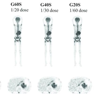
 Imaging of an 11-year-old child with neuroblastoma, with the maximum intensity projection (MIP) of the half-dose image and coronal view and axial images of the serial dose reduction image generated by reduced count. The overall image quality scores of G600 to G20 were 5, 5, 4, 3, 3 and 2 points. The lesion was even identifiable reduced down to a 1/60-dose. Image courtesy of Dr. Wanqi Chen.
The team found image quality scores in the generated 20-second whole-body PET/CT images were significantly lower but that sufficient subjective image quality and lesion conspicuity could be maintained in the other images.
Specifically, the study authors determined that images generated with a 60-second acquisition time with a dose reduction of 0.185 F-18 FDG MBq/kg enabled the identification of 100% of the cancer lesions in the patients, Chen explained.
"Total-body PET/CT with half-dose F-18 FDG achieved good performance in pediatric oncological patients ensuring sufficient image quality and lesion conspicuity," Chen concluded.