Doctors recently used prenatal ultrasound to detect a rare 4.6 x 3.6-cm oropharyngeal teratoma (OPT) protruding from the oral cavity of a fetus. Details of the case were published on November 4 in the Cureus Journal of Medical Science.
Fetal oropharyngeal tumors, which can obstruct breathing, have high morbidity and mortality rates, and early diagnosis can improve patient outcomes. Unfortunately, in this case, the day after imaging occurred, the mother spontaneously delivered the baby at 27 weeks and the child died soon after birth, according to the authors.
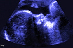
 Ultrasound shows an elongated stalk of tissue with a polypoidal mass protruding from the mouth of the fetus and floating in the amniotic cavity. The mass has mixed solid-cystic tissue layers with areas of calcifications and minimal vascularity. All images courtesy of Narayan et al. Licensed under CC BY-NC 4.0.
"Oropharyngeal teratomas are a rare fetal tumor that can be diagnosed with the use of ultrasounds in utero," wrote the authors, led by Dr. Ruchika Narayan from Narayan Medical College and Hospital in Sasaram, India.
Oropharyngeal teratomas occur in 1 in 35,000 to 200,000 live births, according to the authors. With this type of teratoma, tumor cells fill the oral cavity, often resulting in severe obstruction of the upper airways and high rates of death during the neonatal period. It also usually presents with an excess of amniotic fluid.
Oropharyngeal teratomas used to be identified in fetuses very late or even after birth; however, with advances in ultrasonography, these teratomas now can be diagnosed prenatally. MRI also can provide detailed characterization of the lesion and exclude central nervous system involvement and determine the tracheal anatomy for airway safety.
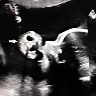
 Sagittal MRI scan shows a mixed-intensity mass with a stalk protruding through the jaws into the amniotic fluid.
A 21-year-old pregnant woman
The woman, who was in her 27th week of pregnancy, visited the hospital because she had lower abdominal pain and increasing stomach girth. She had no significant medical history or family history of hereditary disease or malformations. However, clinicians suspected the woman had excess amniotic fluid.
An ultrasound revealed a pedunculated 4.6 x 3.6-cm polypoidal mass protruding from the mouth of the fetus and freely floating in the amniotic cavity. The mass had a mixed solid-cystic echotexture, and it showed areas of calcifications and minimal vascularity. The woman also had an elevated level of amniotic fluid, with an amniotic fluid index greater than 28.5, according to the authors.
An MRI scan was performed to obtain better characterization of the tumor and to evaluate the association between the mass and surrounding structures. The MR images revealed a mixed-intensity mass with a stalk protruding through the jaws into the amniotic fluid. The internal part of the mass started at the hard palate and filled a large part of the oropharynx. The mass wasn't associated with any central nervous system anomaly, the authors wrote.
 The fetus and mass protruding from the oral cavity.
A day after imaging, the patient had spontaneous delivery of a live fetus. The previously identified mass was large, soft, and mobile with surface hemorrhaging and was protruding from the oral cavity. The baby died of severe respiratory distress shortly after birth before doctors could intervene. Histological analysis confirmed the mass was a teratoma, according to the authors.
Brighter outcomes in the future
The woman is now in the third trimester of a new pregnancy. So far, no fetal abnormalities have been detected. The authors reviewed 33 cases of fetuses with oropharyngeal teratomas, and of these, 12 fetuses died in utero or shortly after birth. Narayan and colleagues noted that they are unaware of any reported cases of recurrence of oropharyngeal teratomas in subsequent pregnancies.
"Early diagnosis of OPT is of great value for obstetric and neonatal management," they added.