MRI scans of patients with traumatic head injuries have revealed microbleeds that appear as dark lesions and are often too small for detection on CT, according to a study published on October 14 in Brain.
Researchers from the U.S. National Institutes of Health's National Institute of Neurological Disorders and Stroke (NINDS) even found evidence of microbleeds among patients with mild traumatic head injuries and a trend toward worse outcomes for patients with the anomalies.
"Traumatic microbleeds may represent injury to blood vessels that occur following even minor head injury," said senior study author Lawrence Latour, PhD, NINDS researcher in a statement. "While we know that damage to brain cells can be devastating, the exact impact of this vascular injury following head trauma is uncertain and requires further study."
Microbleeds essentially are small cerebral hemorrhages caused by structural damage to small vessels in the brain. The detection of these microbleeds after head trauma is particularly critical, since the abnormalities can lead to cognitive and memory decline and have been linked to neurodegeneration, stroke, and Alzheimer's disease.
To determine the connection between traumatic brain injury, microbleeds, and patient outcome, Latour and colleagues from the Cold Spring Harbor Laboratory and the Uniformed Services University of the Health Sciences enrolled 439 adults who experienced head injury and were treated in the emergency department. The patients underwent MRI scans within 48 hours of the injury and were imaged again during four follow-up visits. The subjects were also asked to complete behavioral and outcome questionnaires to chart postinjury symptoms and their progress.
Brain MR images showed evidence of microbleeds in 58% of patients with severe head injury. Most interestingly, 27% of patients with mild cases of head injury also presented with microbleeds on MRI.
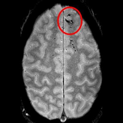
 Traumatic microbleeds appear as dark lesions on MRI scans and suggest damage to brain blood vessels after head injury. Image courtesy of Latour Lab/NINDS.
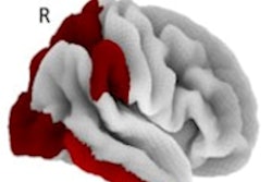
The MR images showed the microbleeds as either linear streaks or as dotted lesions, both of which were observed in a majority of patients. The most common location for the microbleeds was in the frontal lobes of the patients, which would indicate head trauma near the forehead. Patients with microbleeds were also more likely to have moderate to severe disability than patients without microbleeds.
In addition, Latour and colleagues performed a postmortem MRI scan and histological analysis of one study participant's brain that was donated by the family for further evaluation. The scan and tests revealed iron deposits, indicating blood in the brain's immune cells. This finding allowed the researchers to track the effects of the microbleeds beyond initial and follow-up MRI scans.
"Combining these technologies and methods allowed us to get a much more detailed look at microbleed structure and get a better sense of just how extensive they are," added first author and graduate student Allison Griffin.
The authors concluded that the presence of microbleeds after a traumatic brain injury potentially could become a biomarker for identifying which patients are likely to benefit from vascular treatments. They also recommended additional research to determine the aftereffects of microbleeds and the best strategies to treat them.
In the meantime, Latour and colleagues added that there is currently no evidence that MRI scans should replace CT scans for suspected head injury.