A near-infrared imaging technology may be able to identify small cancers by tracking fluorescent probes deep in the body, according to research published March 7 in Scientific Reports.
Imaging technology such as CT and MRI can image the whole body but can't reliably identify tumors until they reach about 1 cm in size, according to a team led by Xiangnan Dang, PhD, of the Massachusetts Institute of Technology (MIT). Working with Angela Belcher, the James Mason Crafts Professor of Biological Engineering and Materials Science and Engineering at MIT, Dang's group sought to develop technology that could image small groups of cells deep within tissue without radioactive labeling.
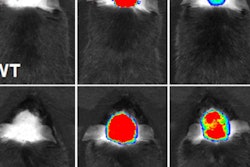
Dang's team conducted an animal study in mice with the imaging system, called DOLPHIN (detection of optically luminescent probes using hyperspectral and diffuse imaging in near-infrared), using it to track a 0.1-mm fluorescent probe through the animals' digestive tracts. The group discovered that the system could detect a signal to a tissue depth of 8 cm.
"In terms of practical applications, this technique would allow us to noninvasively track a 0.1-millimeter-sized fluorescently labeled tumor, which is a cluster of about a few hundred cells," said study co-author Neelkanth Bardhan, PhD, in a statement released by MIT. "To our knowledge, no one has been able to do this previously using optical imaging technique."
Going forward, the researchers are using a related version of the system to try to detect ovarian cancer and have begun working on adapting the technology to identify pancreatic cancer, brain cancer, and melanoma.