Using functional MRI (fMRI), researchers detected changes in brain activity that could relate to one's ability to lose weight. Results of the study were published online October 18 in Cell Metabolism.
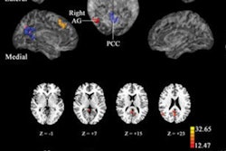
The researchers scanned 24 people at a weight loss clinic before starting them on a standard weight loss diet of 1,200 kcal/day. The fMRI scans targeted the lateral prefrontal cortex, which is associated with self-regulation, and the ventromedial prefrontal cortex, which is involved in motivation, desire, and value.
Subjects were shown pictures of appetizing foods as well as pictures of scenery, which served as the control. Comparisons of brain-activity response to the food pictures, particularly the high-calorie food pictures, were performed for each subject at baseline, one month, and three months. When the subjects viewed appetizing food photos, the ventromedial prefrontal cortex area became more active on fMRI.
Regarding weight loss, at one and three months, the signal from the ventromedial prefrontal cortex decreased, and it dropped the most in people who were more successful at losing weight. In addition, activity in the lateral prefrontal cortex increased.
"In the fMRI, the self-control area increased its activity and the value area decreased its activity," said study co-author Alain Dagher from the Montreal Neurological Institute and Hospital in Canada. "The amount of change was predictive of successful weight loss."
The results suggest that weight loss treatments that increase self-control may be most helpful, particularly when stress leads to overeating, Dagher added.