Functional MRI (fMRI) offers clues as to which memory lapses in older adults are part of normal aging and which may indicate early stages of diseases such as Alzheimer's, according to research published online March 7 in Neuron.
A team led by Zachariah Reagh, PhD, from the University of California, Davis found that data from high-resolution brain fMRI showed some of the underlying causes for differences in memory proficiency between older and younger adults. The study included 20 young adults (ages 18 to 31) and 20 cognitively healthy older adults (ages 64 to 89) who were asked to perform an object memory task and a location memory task while undergoing an fMRI exam.
In the object task, participants were shown pictures of everyday objects and asked to distinguish them from additional pictures, some of which were identical, some of which were new, and some of which were similar but with differences in color or size. The second task required participants to determine whether the objects had changed location.
Object memory is more vulnerable than spatial memory in the early stages of brain diseases such as Alzheimer's, the group wrote. By scanning study participants' brains while they underwent fMRI, Reagh and colleagues were able to establish a mechanism within the brain for that deficit in object memory, finding that it was linked to a loss of signaling in the anterolateral entorhinal cortex. The researchers did not find age-related differences in another area of the brain connected to memory, the posteromedial entorhinal cortex, which supports spatial memory function.
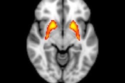
 The figure shows two different brains aligned to a common template space for comparison. The yellow in the anterolateral entorhinal cortex of the young brain indicates significant activity, something that is absent in the older brain. Courtesy of Zachariah Reagh, PhD.
Reagh and colleagues now plan to explore whether this type of fMRI scan could eventually be used as a tool for early diagnosis. They will expand their work to a sample of 150 older adults who will be followed over time, and the subjects will also receive PET scans to track amyloid and tau pathology.