As a medical physicist at a large public trauma hospital in Melbourne, Australia, I'm not a frontline worker, but many of my colleagues in radiology are face-to-face with COVID-19 positive or suspected patients. As staff get more fatigued and confront the many challenges of the pandemic, I wanted to support them by making their work safer and easier.
We heard about a clever technique for taking chest x-rays through glass. In theory, this allows the x-ray unit to remain outside an isolated patient's room -- conserving personal protective equipment (PPE), reducing radiographer risk, and speeding up the process.
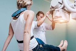
 The radiographer inside the cubicle positions the bed close to the glass door and places the digital detector behind the patient for an erect anterior-posterior chest x-ray before stepping away from the patient during the exposure. The radiographer outside the cubicle positions the x-ray tube head close to the glass door and steps laterally away to initiate the x-ray while maintaining visual contact with the patient. The subject in the figure is not a patient but the author of this publication. Reproduced with permission of the Australasian College of Physical Scientists and Engineers in Medicine (ACPSEM).
We first saw details about the technique on Twitter. Also, authors of a 2016 article have explained about imaging and the infection control aspects for the Ebola virus,1 which seemed equally applicable to COVID-19.
The first step was to convince our staff this was a safe and reasonable way to take chest x-rays, but it was not easy to find publications that explained the radiation safety or image quality aspects of the technique. This motivated us to answer questions about the magnitude of x-ray transmission through glass, safety aspects due to scattered radiation, and the technique parameters that would be most useful.
We've published our findings recently in Physical and Engineering Sciences in Medicine.2 We found that 110 kV and 5.5 mAs were the most commonly used factors to account for the glass and additional distance to the patient (this could be up to 3 m between the x-ray tube and detector). The glass was typically equivalent to about one half-value-layer (i.e., it reduced the intensity of the x-ray beam by about 50% and also increased the beam quality or average energy of the x-ray beam).
Our radiologists were satisfied with the image quality, with 90% of images taken through glass considered diagnostic. With our parameters, the typical radiation dose to patients is the same whether the x-ray is taken through glass or not.
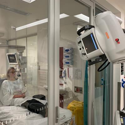
 An example of how the technique can obtain good image quality (110 kV, 5 mAs). Reproduced with permission of ACPSEM.
The technique has been established for use in the current COVID-19 pandemic, and we assessed the radiation safety accordingly. The radiographers and/or nurses do not need to wear a lead apron. This was important because of the amount of PPE they were already wearing and the infection control risks associated with sharing lead aprons.
The radiation dose to staff standing one meter away while a chest x-ray is taken through glass is equivalent to approximately three hours of natural background radiation in Australia (less than 0.5 microsieverts, µSv) and even lower further away. We encourage staff to maximize their distance from the patient being imaged and also from the backscatter off the glass outside the room.
We have now performed several hundred chest x-rays using this technique. In intensive care unit (ICU) rounds, one radiographer and a nurse tend to stay in the room, while the second radiographer operates the x-ray unit from outside the room. In the emergency department, where the rooms are not as large, staff leave the room while the x-ray is performed. This technique should never be used through lead glass.
Staff need to consider whether the technique is appropriate for each individual patient and also the clinical question. For example, it's not going to be useful where a large patient is semi-erect to confirm the wide-bore nasogastric tube position. In these cases, a supine in-room technique will answer the clinical question more quickly and with less chance of needing to repeat the x-ray.
 Zoe Brady, PhD.
One of the highlights of implementing this technique was the teamwork and collaborative spirit displayed. Radiologists, radiographers, physicists, infection prevention nurses, ICU nurses, and researchers all worked together. Adaptability is key in our response to the pandemic.
This technique strengthens our defence against COVID‑19. All of us have stretched resources coping with the pandemic and we hope our "how to" guide provides enough information for others to put the chest x-ray through glass technique into practice.
References
- Thompson N, Garrahy P, Olivieri G. Ebola Virus Disease, An Imaging Protocol. Spectrum. Melbourne, Australia: Australian Institute of Radiography, Wiley Blackwell; 2016;23(16):18-21.
- Brady Z, Scoullar H, Grinsted B, et al. Technique, radiation safety and image quality for chest x-ray imaging through glass and in mobile settings during the COVID-19 pandemic. Phys Eng Sci Med. 2020:1-25. doi:10.1007/s13246-020-00899-8.
Zoe Brady, PhD, is chief physicist in radiology and nuclear medicine at Alfred Health, Melbourne, Australia. Competing interests: None declared.
The comments and observations expressed herein do not necessarily reflect the opinions of AuntMinnie.com, nor should they be construed as an endorsement or admonishment of any particular vendor, analyst, industry consultant, or consulting group.