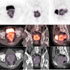
People who have received frequent dental x-rays have an increased risk of developing meningioma, the most commonly diagnosed primary brain tumor in the U.S., according to a study published today in Cancer (April 10, 2012).
The findings should serve as a reminder to patients and practitioners alike to carefully assess the need for diagnostic imaging procedures and to limit the frequency of x-rays based on the recommendations of professional organizations, such as the ADA.
Ionizing radiation is the primary environmental risk factor for developing meningioma, and dental x-rays are the most common artificial source of exposure to ionizing radiation for individuals in the U.S., the study authors noted.
To examine the link between dental x-rays and the risk of developing meningioma, Elizabeth Claus, MD, PhD, of the Yale University School of Medicine and Brigham and Women's Hospital, together with colleagues from multiple other academic medical centers, studied information from 1,433 patients who were diagnosed with the disease between the ages of ages 20 and 79 and were residents of the states of Connecticut, Massachusetts, North Carolina, the San Francisco Bay Area, and eight counties in Houston, TX, between May 1, 2006 and April 28, 2011.
They also studied information from a control group of 1,350 individuals who had similar characteristics but who had not been diagnosed with a meningioma.
To avoid attributing the effect of therapeutic ionizing radiation to dental x-rays, individuals who had received therapeutic radiation to the head, neck, chest, or face were removed from all analyses that assessed the risk associated with dental x-rays, the study authors noted.
"All epidemiology studies are imperfect and only suggest a correlation, not a cause and effect," Dr. Claus said. "But we did collect information on other sources of common ionizing radiation, particularly therapeutic. We tried to control for other things that we knew would be an issue."
'Apparent association'
Over a lifetime, patients with meningioma were more than twice as likely as controls to report having ever had a bitewing exam, the researchers found. Individuals who reported receiving bitewing exams on a yearly or more frequent basis were 1.4 to 1.9 times as likely to develop meningioma as controls. (Risks differed depending on the age at which the exams were done).
An increased risk of meningioma was also linked to panorex exams taken at a young age or on a yearly or more frequent basis. Individuals who reported receiving these exams when they were younger than 10 years old had an increased risk (4.9 times) of developing meningioma. Those who reported receiving them on a yearly or more frequent basis were 2.7 to 3 times (depending on age) as likely to develop meningioma as controls.
Previous studies have reported similar findings, the study authors noted (Neuroepidemiology, 1989, Vol. 8:6, pp. 283-295; Oral Oncology, July 1998, Vol. 34:4, pp. 265-269; Cancer, March 2004, Vol. 100:5, pp. 1026-1034). However, no recent large-scale studies of meningioma risk relative to common ionizing radiation exposure exist, when doses for dental and other procedures have decreased but new radiography procedures have been introduced, they added.
"To our knowledge, this is the largest case-control study to date examining the correlation between dental x-rays and the risk of meningioma," the study authors wrote. "And because it is the most recent study, it provides an improved examination of the effects of reduced dosing exposure levels over time."
Dental patients today are exposed to lower doses of radiation than in the past, they noted. Allan Farman, BDS, MBA, PhD, DSc, a professor of radiology and imaging science at the University of Louisville in Kentucky and immediate past-president of the American Association for Oral and Maxillofacial Radiology, agreed.
"This is not the first time that the subjective memories of patients with meningioma have resulted in an apparent association between dental x-ray use and the likelihood of meningioma," Dr. Farman told DrBicuspid.com. "In each case, the conclusions clearly state that associations are related to radiation received many years ago when dental x-ray exposures were substantially higher than is presently the case.
"Perhaps inclusion of information on radiation exposure of non-dental origin, such as medical CT to the head -- which is orders of magnitude greater in dose than that from dental radiographs -- would have been beneficial to better view the findings in a wider context."
Time to 'Image Wisely'
Other study limitations include the possibility of under- or over-reporting of dental x-rays by the participants, Dr. Claus and her co-authors noted.
"This is a difficult problem in epidemiology because, unlike medical care, which ... may be confirmed by a review of centralized medical records, dental care generally is obtained ... from numerous dentists, all of which are outside of a health maintenance organization or hospital-based setting, providing little opportunity for researchers to validate dental reports in a timely or cost-efficient manner," they wrote.
Arthur Goren, past director of radiology at SUNY Stony Brook School of Dental Medicine and currently clinical associate professor in the department of cariology and comprehensive care at New York University College of Dentistry, also questioned the study's methodology.
"The only way bitewing x-rays can cause brain tumors is through scatter radiation," he told DrBicuspid.com. "If the scatter reaches the brain, why are tumors not found in the thyroid, eye, parotid, and oral epithelium? We have dosimetry studies of cone-beam CT exposures to the head and neck of adult male and female and juvenile phantoms that show that the other organs received more scatter radiation than the brain and cranium.
"I realize CBCT dosimetry is not an adequate comparison with bitewing dosimetry, but to base findings on anecdotal recollections and not on statistical data is a reach," he added. "I wonder what the conclusions would be if you substituted cell phone use instead of bitewing x-rays."
Nevertheless, Dr. Farman said, "this study has value in that it does remind clinicians (including dental practitioners) that x-radiation has been defined by the U.S. Food and Drug Administration as a carcinogen and that radiographs should only be made following professional prescription and never as a mere routine."
The study presents an ideal opportunity in public health to increase awareness regarding the optimal use of dental x-rays, which, unlike many risk factors, is modifiable, Dr. Claus noted. The ADA's guidelines for healthy persons suggest that children receive 1 x-ray every one to two years; teens receive one x-ray every one-and-a-half to three years, and adults receive one x-ray every two to three years (Journal of the American Dental Association, September 2006, Vol. 137:9, pp. 1304-1312).
The use of ionizing radiation should always be based upon professional judgment after taking a history and clinical inspection, Dr. Farman added.
"We should always 'image wisely' irrespective of the region being examined, and this includes radiographs needed for dental diagnosis," he said. "With careful selection made by a professional, there is no doubt that dental-imaging procedures can reduce pain and suffering and also that early intervention can reduce the costs of treatment intervention involved."