Applying morphologic techniques to teeth to calculate a person's age requires extracting at least one tooth. But that isn't always possible, particularly if the individual in question is still alive (and even if he or she is dead, sometimes religious or cultural beliefs prevent body parts from being removed).
In the mid-90s, Dr. Sigrid Kvaal and colleagues in Oslo, Norway, proposed x-ray as a noninvasive way to estimate the age of adults in forensic work and archaeological studies (Forensic Science International, July 28, 1995, Vol. 74:3, pp. 175-185).
More recently, a group from the Katholieke Universiteit in Leuven, Belgium, tested Kvaal's dental age calculation technique on panoramic dental radiographs. Dr. Nathalie Bosmans and colleagues gathered 197 digital orthopantomographs from three Belgian dental practices. The subjects were all Caucasian (95 females, 102 males) with a mean age of 34.7 years.
Six teeth were selected for analysis per Kvaal's method:
- Maxillary central
- Lateral incisors
- Second bicuspids
- Mandibular lateral incisors
- Canines
- First bicuspids
For all six teeth, Bosmans' group measured the maximum tooth length, the pulp length, the root length on the mesial surface from the enamel-cementum junction (ECJ) to the root apex, the root and pulp width at the ECJ level, the midroot level, and the midpoint between the ECJ and the midroot level. Software that automatically calculates dental age was used to analyze the data. Months later a single observer evaluated the x-rays.
 |
| "Morphologic assessment is great in dead individuals. In living people, you only have the radiology option," co-author Dr. Guy Willems said. The observer in this study had no idea as to the chronological age of the individuals whose panoramic x-rays were used. |
"Statistical analysis was carried out in order to spot significant differences between the chronological age and the calculated age," the authors explained (Forensic Science International, October 29, 2005, Vol. 153:2-3, pp. 208-212).
The results showed a high concordance correlation between the calculated dental age and the chronological age when measurements from all six teeth were used or when only three mandibular teeth were used. However, using any other combination of teeth resulted in significant differences the between calculated age and the real age.
The authors concluded that their high degree of intraobserver agreement indicated the high reproducibility of using Kvaal calculation on panoramic x-ray.
 |
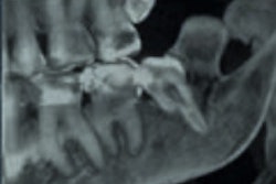
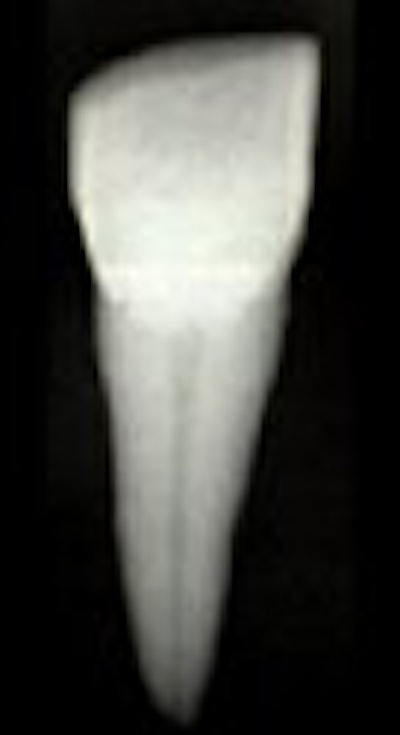
| Kvaal's technique examines the relationship between chronological age and the 2D dental pulp size, including length and width, in individuals older than 20. Images courtesy of Dr. Guy Willems. |
In an e-mail interview with AuntMinnie.com, co-author Dr. Guy Willems -- who, along with Bosmans, is from the university's department of forensic odontology -- explained that panoramic x-rays offer some advantages over standard, long-cone periapical radiographs.
"(Panoramic x-rays) are more commonly taken, and it would take only this (one) radiograph to visualize all the teeth you need," he said.
By Shalmali Pal
AuntMinnie.com staff writer
October 12, 2005
Related Reading
Bone loss puts older women at risk of tooth loss, August 5, 2005
Sophisticated 3D volumetric imaging has a place in everyday orthodontics, July 4, 2005
CT helps unwrap mummy mystery, May 29, 2005
Radiologic 'autopsy' is already finding evidence without destroying it, December 4, 2003
Copyright © 2005 AuntMinnie.com