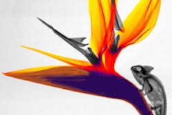
Radiography has traditionally been used to examine the internal structure of muscles, organs, and bones, but a few medical imaging specialists have gone a step further, turning their images into art. Steven Meyers is one of those people. His Web site, www.xray-art.com, exposes his inner vision of organic structure through x-ray images of flowers.
Meyers, a radiology technologist for 30 years and a self-taught landscape photographer for 23 years in the Seattle area, hopes to revive floral radiography as an art form.
 |
"I'm on a mission to get x-ray art seen and to establish it as legitimate art. I haven't seen any new floral radiographs in the last 20 years or so, and there are only a few radiographic artists in the world. It is my intention to keep the art form alive with new and exciting images," Meyers said.
Meyers creates his images using a Micron Focus 50 specimen radiography system with a focal spot of 0.05 mm, variable kilovoltage (10-40 kVp), and fixed milliamperage. The radiography cabinet, which he said has been discontinued but is similar to the Faxitron MX-20, is commonly used in radiology and pathology labs to make radiographical images of biopsy and surgical specimens.
 |
"The system provides a soft x-ray that's ideal for floral subjects. Most standard hospital equipment is too powerful for floral radiographs," he said.
His preferred film is the Min-R 2000 mammography product (Eastman Kodak, Rochester, NY), which he exposes directly on 8 x 10-inch sheets. Meyers said the most difficult aspect of creating floral radiographs is attaining the perfect composition.
"I'm on a constant quest for the perfect radiograph -- composition is everything. Of the 1,200 images I've made with this technique, 95% fail because of composition. There's no lens to look through to compose images, and [not] all floral subjects make interesting radiographs. Many floral subjects that make spectacular photos make surprisingly uninteresting radiographs," he said.
 |
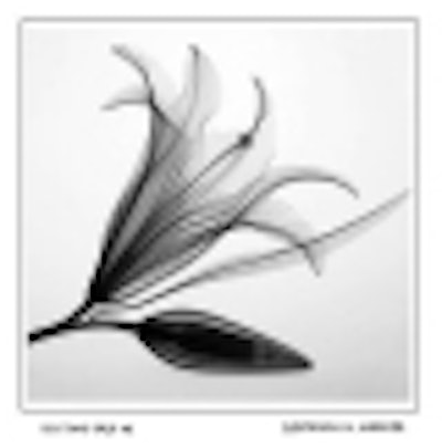
Meyers uses the anode-heel effect to produce the light-to-dark gradient background. The effect, he said, is more prominent at low exposures. To enhance the texture and detail of the stem and petals, he uses negative, positive, and solarized images.
"Since the technique is unnatural by most standards, I try to emphasize the unnatural parts [of floral structure] in the final prints," he said.
Once he settles on the perfect composition, Meyers acquires a high-resolution digital scan of the image and uploads the data to a computer to make adjustments. He then outputs the data to a digital film recorder to attain a 4 x 5-inch black and white negative. Finally, he enlarges the image onto fine art silver-rich paper. Meyers' darkroom prints are limited to 50.
He will soon only offer quadtone ink prints of his floral radiographs. With this technique, he creates a high-resolution digital scan of the original image and sends it directly to his quadtone printer, which is loaded with carbon pigment inks in black and three shades of gray.
"The quadtone prints are sharper and more detailed. It looks fantastic, especially on images that resemble drawings," he said.
 |
Dr. Dain Tasker first introduced floral radiography in the mid-1930s. Born in 1872 in Beloit, WI, Tasker was the chief radiologist at Wilshire Hospital in Los Angeles when radiology was in its formative years.
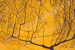
Tasker chose not to "pose" or "chemicalize" his floral subjects. He used weak iodine solutions to naturally expose the "circulatory system of the stem and petals" (Naked to the Bone: Medical Imaging in the 20th Century, Bettyann Holtzmann Kelves, Addison-Wesley, Reading, MA 1997).
"When we eliminate color and fall back on outline and molded density as the basic beauty factors we begin to realize nature's infinite variety of structural perfections," Tasker said.
Meyers said his interests aren't limited to flowers. He hopes to radiograph other subjects in nature as well.
"There are things I still very much want to do with x-ray art -- more botanical studies and maybe some other objects. There are lots of possibilities, but I would probably have trouble selling insect pictures," he said.
By Jennifer LungAuntMinnie.com staff writer
July 17, 2001
Click here to post your comments about this story. Please include the headline of the article in your message.
Copyright © 2001 AuntMinnie.com