An experimental chest x-ray method may be useful for measuring air flow in the lungs of patients with chronic obstructive pulmonary disease (COPD), according to a study published January 31 in European Radiology Experimental.
U.S. and Japanese researchers built a prototype vector-field dynamic chest x-ray (VF-DXR) system equipped with optical flow method (OFM) image reconstruction software. They showed that the approach can measure the velocity of airflow in patients with COPD compared with healthy volunteers.
"This current study demonstrated VF-DXR with OFM is applicable to clinical research," wrote corresponding author Dr. Takuya Hino of Harvard University's Center for Pulmonary Functional Imaging in Boston.
Eventually, the method could enable clinicians to simultaneously assess anatomical and physiological information in these patients, as well as reduce cost and radiation exposure associated with traditional CT imaging, according to the authors.
COPD is one of the leading causes of morbidity and mortality worldwide. Patients diagnosed with the condition have decreased mobility of the thorax, which affects their breathing. Pulmonary function tests such as lung volume and forced expiratory volume (FEV1), which measures the amount of air that can be forced from the lungs in one second, are major criteria for assessing the severity of COPD.
Dynamic x-ray is a method first developed by researchers and industry collaborators in 2006. This system transmits a series of pulsed x-rays about 15 times per second using the same principle as animation and displays a series of static images to create dynamic images. Dynamic chest x-ray (DXR) allows researchers to observe actual movement.
Previously, Hino and colleagues showed the efficacy of DXR in measuring diaphragm or rib motion, as well as projected lung area during a particular breathing phase in the standing position and that DXR was able to acquire sequential images with high temporal resolution.
Hino and colleagues have also previously reported changes in diaphragm motion between normal subjects and COPD patients with DXR. In this study, the researchers aimed to assess the quantitative difference of dynamic lung motion between patients with COPD and healthy volunteers using DXR with OFM, an imaging reconstruction method that quantifies movement.
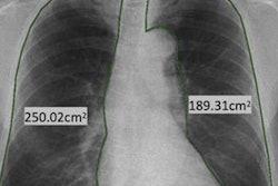
The researchers enrolled 36 patients with mild to severe COPD and 47 healthy volunteers. Posteroanterior view of chest DXR examination in standing position were performed with the prototype x-ray system (Konica Minolta, Japan) using a flat-panel digital detector and a pulsed x-ray generator with cineradiography.
During the examination, COPD patients took several tidal breaths and one forced breath separately, while control subjects took several tidal breaths, followed by one forced breath. The researchers assessed pulmonary function data.
Results confirmed negative correlations between lung motion velocity and the percentage of forced expiratory volume in one second (%FEV1) in the tidal inspiration in the right lung (rs = -0.47, p < 0.001) and the left lung (rs = -0.32, p = 0.033). A positive correlation between lung motion velocity (LMV) and %FEV1 in the tidal expiration was observed only in the right lung (rs = 0.25, p = 0.024).
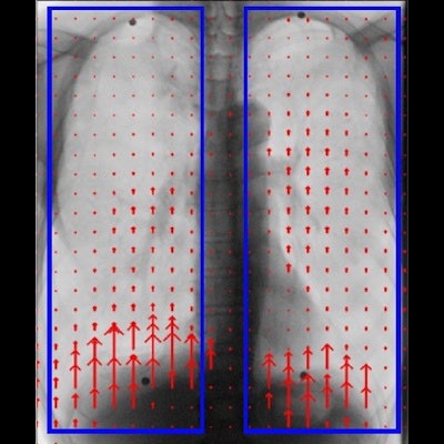
 A 65-year-old male COPD patient with 38.5 FEV1% and percent predicted %FEV1 16.3, which meets the criteria of severe COPD. Blue rectangles (d) represent bilateral regions of interest. VF-DXR images in (a) tidal inspiratory, (b) tidal expiratory, (c) forced inspiratory, and (d) forced expiratory phase. (e) The graph shows the temporal change between LMV in the right lung and the number of frames. The frame rate is set to 5 frames/s (Video 2 in the Supplemental material). Images courtesy of European Radiology Experimental.
Lung motion velocity among groups characterized as being normal, having mild COPD, or having severe COPD were different in the tidal respiration, the researchers stated. The COPD mild group showed a significantly larger magnitude of lung motion volume compared with the normal group, they added.
"In the tidal inspiration, the lung parenchyma moved faster in COPD patients compared with healthy volunteers. VF-DXR was feasible for the assessment of lung parenchyma using [lung motion volume]," the authors stated.
The authors noted that one unique characteristic of the DXR method is that it doesn't require physical contact between the patients' mouth and pulmonary function equipment, which could be helpful against possible emerging infection such as seen in the COVID-19 pandemic or novel respiratory diseases in the future.
The absence of intrinsic circuit resistance is another unique characteristic of the approach, they added.
Yet further investigations are needed to utilize DXR more effectively and to establish it as a valuable diagnostic tool, they concluded.