Three-tesla 3D MR spectroscopy provides high diagnostic sensitivity and specificity for distinguishing between benign and malignant breast lesions within reasonable measurement times, according to an Austrian study published in the December issue of Radiology.
Researchers from the Medical University of Vienna concluded that MR spectroscopy produced a sensitivity of 97% and a specificity of 84%. In addition, based on certain threshold values for choline signal-to-noise (S/N) ratio, 3D MR spectroscopy provided high diagnostic accuracy in 11 minutes and 17 seconds.
The lead author of the study is Stephan Gruber, PhD, from the department of radiology and MR Center of Excellence (Radiology, Vol. 261:3, pp. 752-761).
The prospective study consecutively enrolled 53 female patients with a mean age of 50 years (range, 25-82), who were referred for 3-tesla MRI based on clinically suspicious findings and/or abnormalities detected with mammography and/or ultrasound.
MRI scans were conducted on a 3-tesla system (Magnetom Trio, Siemens Healthcare) in the prone position with a dedicated bilateral breast coil (Invivo). The exams occurred in the second week of a patient's menstrual cycle.
The researchers added 3D MR spectroscopy to the clinical protocol that included a 3D T1-weighted turbo fast low-angle shot (FLASH) sequence, a T2-weighted short-tau inversion recovery (STIR) sequence, and a breath-hold examination sequence before and after contrast agent was administered. MR spectroscopic images were taken before contrast agent administration to avoid any undue influence from the medium on the detected choline signal.
Because of previous mammography and ultrasound results, the researchers knew the approximate location of breast lesions, which were verified by histopathology or at follow-up of at least 24 months.
A total of 32 malignant tumors and 12 benign lesions were confirmed in 43 patients by histopathology. Seven patients without biopsy also received imaging follow-up. The maximum choline S/N ratio values of each lesion were compared with the results obtained with contrast-enhanced MRI.
Choline was detected in 31 (97%) of the 32 malignant tumors and in 10 (53%) of 19 benign lesions. The median choline S/N ratio in malignant lesions was 5.7, compared with 2.0 in benign lesions.
A choline S/N ratio threshold level of 2.6 was determined based on a receiver operator characteristics (ROC) curve, which resulted in sensitivity of 97% (95% confidence interval: 82%-100%) and specificity of 84% (95% confidence interval: 72%-100%). Three benign lesions were classified as false positive.
 |
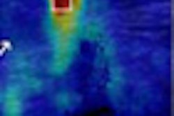
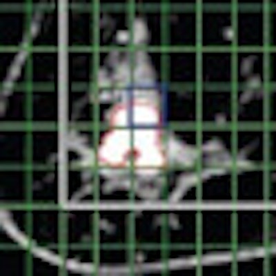
| MR images of invasive ductal carcinoma in a 62-year-old woman. Both images show how shifting MR spectroscopic imaging voxels can be performed retrospectively in all three spatial dimensions to allow improved matching with the pathologic area. Images courtesy of Radiology. |
"Our retrospectively calculated threshold level of 2.6 at 3 tesla indicates that benign lesions may show detectable choline," the authors wrote. "This finding highlights the importance of threshold levels for the determination of diagnostic accuracy."
By comparison, contrast-enhanced MRI showed similar diagnostic accuracy with a sensitivity of 97% (95% confidence interval: 82%-100%) and a specificity of 84% (95% confidence interval: 82%-100%), but with different cases with false-positive and false-negative findings.
Diagnostic accuracy was found to be comparable to that of other MRI methods used in breast imaging, in addition to being comparable to the contrast-enhanced MRI results in this study.
Furthermore, the combination of threshold values for choline S/N ratio and 3D MR spectroscopy produced the highly accurate diagnostic results in 11 minutes and 17 seconds.
Future studies should focus on the value of 3D MR spectroscopy as part of a multiparametric MR protocol that includes diffusion-weighted imaging and contrast-enhanced MRI, Gruber and colleagues recommended.
"We also found a significant difference in choline [signal-to-noise ratio] between low-grade and high-grade lesions," the authors wrote. "This was, to our knowledge, not previously observed with MR spectroscopy, but similar findings were recently reported for diffusion-weighted imaging of the breast."