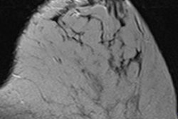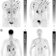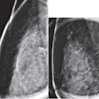
Although it's still an investigational modality, dedicated breast CT offers distinct benefits for diagnostic workup compared to other imaging modalities, according to an article published online in the Breast Journal. Researchers believe that breast CT could handle a wide range of breast imaging tasks, from diagnostic to adjunctive screening applications.
Ultrasound and digital breast tomosynthesis offer advantages when used with screening mammography, and they are relatively easy to incorporate into clinical practice; however, they have limits for diagnostic workup that could be addressed by the judicious use of dedicated breast CT, argue the group from the University of Rochester in New York and the University of Massachusetts in Worcester.
"Tomosynthesis is all the rage, and it's easy to add to a screening protocol," lead author Dr. Avice O'Connell told AuntMinnie.com. "But I see dedicated breast CT as offering a technological advantage after mammography and tomosynthesis for workup, because it delivers true 3D images."
Mammography's limitations
Mammography's sensitivity limitations, particularly in dense breasts, have led to the use of additional screening modalities such as whole-breast ultrasound, breast-specific gamma imaging, and digital breast tomosynthesis -- especially as more states pass density notification laws. But dedicated breast CT for follow-up could reduce the need for other techniques (Breast Journal, September 8, 2014).
 Dr. Avice O'Connell from the University of Rochester.
"As the initial finding usually originates from the mammogram, it is natural that the radiologist would like the benefit of visualizing this finding with better contrast and in 3D detail that demonstrates its relationship with the surrounding anatomy," O'Connell and colleagues wrote. "Dedicated breast CT is capable of providing increased contrast and fully 3D anatomic information."
Clinicians have long been interested in using CT to image the breast, but conventional CT scanners send x-rays through the entire thorax, which raised concerns about patients' radiation exposure, according to the authors. In recent years, CT technology has improved, leading to the development of dedicated conebeam systems such as the investigational Koning unit that O'Connell and colleagues used. (Koning received the CE Mark for the device in 2012 and Canadian regulatory approval earlier this year.)
Breast CT's benefits
Dedicated breast CT for diagnostic workup has some key benefits compared to other technologies, according to O'Connell and colleagues.
No compression
Dedicated breast CT doesn't require compression and it produces images that are not compromised by the tissue overlap inherent in mammography, O'Connell and colleagues wrote. Breast MR and automated whole-breast ultrasound also avoid tissue compression and offer 3D images, but dedicated breast CT provides higher spatial resolution than these other technologies.
Higher slice sensitivity
Tomosynthesis acquires data that are reconstructed into slices to provide a "quasitomographic" view of the breast, the authors wrote. This is helpful, but the data are limited compared to 3D breast CT, in that the tomographic system's ability to separate the slices (its "slice sensitivity profile") worsens with larger breast diameters. Breast CT's slice sensitivity remains constant, and it generates images across any desired plane with consistent resolution.
Shorter acquisition times
Compared with breast MRI, breast CT has shorter image acquisition times: 10 seconds for a complete scan versus four to eight minutes per sequence in MRI. Breast CT may not replace breast MR in a diagnostic setting, but it could be an alternative when access to MR is limited, according to O'Connell's and colleagues.
Addresses several breast density concerns
As more states pass breast density notification laws, the demand for supplemental imaging -- usually whole-breast ultrasound or tomosynthesis -- increases. Compared with ultrasound, breast CT images are more similar to those from mammography, and breast CT can be interpreted on the same image processing and display platform as body CT. It's also less operator dependent, offers the option to biopsy a finding using the modality on which it was found, and can even quantify breast density, the authors wrote.
Radiation dose is reasonable
The low end of the reported average glandular radiation dose from breast CT is similar to the dose from a two-view screening mammography exam and slightly lower than the dose from a combined tomosynthesis-mammography two-view screening exam, they wrote.
The bottom line? Dedicated breast CT has the potential to be better than conventional mammography for the diagnostic evaluation of suspicious findings, O'Connell concluded.
"Having been in the business of breast imaging for 30 years, you get a perspective on what works and what doesn't," she told AuntMinnie.com. "Given all the roles in breast imaging CT could have -- from adjunct screening and diagnostic evaluation, guiding biopsies, evaluating the extent of disease, and even monitoring patients' therapy response -- the technology is poised to become an important imaging tool."
