Access to onsite pathology evaluation can significantly improve the success rate of ultrasound-guided fine-needle aspiration (FNA) biopsy of thyroid nodules, according to research from the University of North Carolina (UNC) at Chapel Hill.
Compared to a patient group evaluated without the presence of an onsite pathologist, onsite pathology evaluation of specimens at the time of biopsy reduced the inadequacy rate by 63%, according to the UNC study team. This improvement was achieved even though the mean nodule size was larger in the patient group without an onsite pathologist.
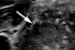
Ultrasound-guided FNA is the standard procedure for characterizing thyroid nodules, and practices are increasingly performing thyroid nodule biopsies. However, the reported adequacy rate for thyroid FNA varies widely due to its subjective nature, according to Wui Chong, MD.
"An inadequate specimen requires a repeat procedure, so anything that can reduce the inadequacy rate for thyroid FNA is desirable," he said.
Chong presented the team's findings during a scientific session at the American Institute of Ultrasound in Medicine (AIUM) annual meeting earlier this year in San Diego.
To determine if having onsite pathology evaluation of specimens at biopsy improves the adequacy rate, the researchers performed a retrospective review of ultrasound-guided FNA of thyroid nodules performed at UNC between 2007 and 2009.
In the first patient group, 200 consecutive ultrasound-guided FNAs were performed by radiologists with an attending pathologist present during the procedure. The pathologist inspected the specimen from each pass as it was obtained for adequacy.
Procedures were performed by radiology residents, fellows, and attending physicians. The slide was prepared by cytopathology technologists, and the number of passes depended on the feedback from the pathologist (range, 2-8 per nodule).
In a second patient group, 200 consecutive ultrasound-guided FNAs were performed by an offsite outpatient center by endocrinologists without a pathologist present for onsite specimen evaluation. Procedures were performed by endocrinology attending physicians and fellows, Chong said. Slides were prepared by the endocrinologists themselves, with three passes made for each nodule.
For all patients, 23-gauge heparinized needles were used with an aspiration technique. Both groups had many years of experience with ultrasound guidance, Chong said. The radiology group used a Sequoia ultrasound scanner (Siemens Healthcare, Malvern, PA), while an Aspen ultrasound scanner (Siemens) was used by the endocrinologists.
The only variables between the two groups were the presence of an onsite pathologist and the number of passes (fixed versus variable). Slides were interpreted by the same cytopathology group using the same criteria: benign, malignant, indeterminate follicular neoplasm, or inadequate.
Adequacy criteria at UNC include six groups of cells of at least 10 epithelial cells each, presence of colloid, atypia, or macrophages in a cystic lesion.
In the group without an onsite pathologist, 13.5% of the FNAs were inadequate, compared with 5% in the group with an onsite pathologist (p = 0.0051).
"This was despite the fact that the mean nodule size in the [onsite pathologist] group was smaller," he said.
The mean nodule size was 2.4 cm for the patient group with an onsite pathologist, compared with a mean nodule size of 2.8 cm for the group without one.
The median number of passes was three in both groups. No significant differences in patient demographics or cytopathological diagnoses were found between the two groups.
Chong acknowledged possible limitations of the study, including the use of real-time guidance by the radiologists and a "mark-and-stick" approach by endocrinologists. While "mark and stick" may not be as effective for ensuring sample accuracy, it should not affect sample adequacy, he said.
Also, slides in the offsite group were prepared by physicians and not trained cytotechnologists, raising the possibility that the inadequacy rate might reflect quality of slide preparation rather than feedback from the cytopathologist. Since 2007, however, endocrinology attendings and fellows have received thorough hands-on training in slide preparation, Chong said.
"I think that both of these possible limitations are less important than they seem," he said.
Chong also noted that CPT code 88172 provides reimbursement for an immediate cytohistologic study to determine the adequacy of specimens at the time of the FNA procedure.
Assessment must be performed by a cytopathologist; it cannot be billed if a cytotechnologist performs an adequacy assessment or prepares the specimen in the absence of a pathologist. It also includes a technical and professional component, and can be used as many times as a pathologist is asked to assess adequacy, Chong said.
"So if we do three passes and three aspirations, and the pathologist is asked to assess three separate passes, 88172 can be billed three times," Chong said.
By Erik L. Ridley
AuntMinnie.com staff writer
July 8, 2010
Related Reading
Calcifications on CT distinguish malignant thyroid lesions, May 12, 2010
Thyroid elastography may reduce unnecessary FNA biopsies, April 21, 2010
New ultrasound techniques show promise in thyroid nodules, February 5, 2010
Risk of thyroid cancer increased in childhood cancer survivors, October 28, 2009
ARRS study: US can help avoid thyroid biopsy, April 23, 2009
Copyright © 2010 AuntMinnie.com