For the first time, researchers have shown that whole-body PET/MR can detect hypermetabolic lesions in cancer patients with similar sensitivity to PET/CT, setting the foundation for future studies to validate the use of PET/MR for specific clinical indications.
Researchers found a correlation between the two modalities despite the different attenuation-correction approaches used: MR-based attenuation correction for MR/PET and CT-based attenuation correction for PET/CT. The group also found comparable tracer-uptake ratios in tumor lesions, further confirming diagnostic similarities between the technologies.
The study was presented at the RSNA 2011 meeting by lead author Dr. Alexander Drzezga, a visiting assistant professor at Harvard Medical School and assistant in neuroscience at Massachusetts General Hospital.
PET/MRI's promise
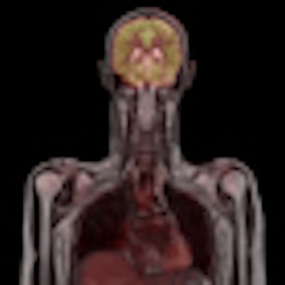
 Whole-body image obtained with Biograph mMR PET/MR system. Image courtesy of Siemens.
PET/MRI is medical imaging's newest hybrid modality, and the first PET/MRI scanners began to receive regulatory clearance in the U.S. this year. Much of the excitement being generated by the technology has to do with the use of MRI rather than CT in combination with PET. In addition to being free of ionizing radiation, MRI has spatial resolution that exceeds CT in many clinical areas.
However, there are still some unknowns surrounding PET/MRI, such as the degree to which PET/MRI images might correlate with PET/CT. Also, PET/MRI scanners use MR images for attenuation correction, which is a different approach than the CT-based attenuation correction to which PET/CT users have become accustomed.
To examine these issues, a total of 32 patients with various oncological diagnoses were evaluated using both imaging modalities. Researchers used a commercially available PET/MR scanner that combines a 3-tesla MRI system with a PET imaging insert inside the bore (Biograph mMR, Siemens Healthcare).
The imaging protocol consisted of a dual-acquisition process, whereby patients underwent the PET/CT scan first and then were subsequently placed on the PET/MR scanner with no additional use of contrast agent. Attenuation correction and regional lesion allocation were performed with low-dose CT for the PET/CT data, and the Dixon MR sequence was used for PET/MRI. The Dixon MR sequence "consists of four different images for attenuation correction of the PET data," Drzezga noted.
For the visual evaluation of images, the study relied on two teams of observers, each consisting of a board-certified radiologist and a board-certified nuclear medicine physician. The radiologist and nuclear medicine physician each subjectively rated the number of suspicious lesions in the PET data, image quality alignment between PET and the contrast of the lesion, and the locations of the lesions based on the PET, CT, and MRI information.
The researchers also based their evaluations on standardized uptake values (SUVs) of the lesions that were detected in the different organs, and they established correlations between the SUVs in both modalities.
'No significant difference'
The visual analysis of the images showed "no significant difference in the number of lesion-positive patients or in the total number of lesions detected in both modalities, which is quite reassuring," Drzezga said. "Also, subjective lesion contrast alignment and anatomic location were comparable" between the two modalities.
"We had slightly lower image quality in the PET/MR data, which was a bit unexpected, but it was basically due to inhomogeneous liver uptake, which had been reported in some of the subjects by the reviewers," he added.
In a quantitative analysis, the researchers found a slight decrease in the mean SUV in lesions between PET/CT and PET/MR, but the difference was not significant. The mean SUV uptake ratio in tumor lesions was 6.4 for PET/CT and 7.3 for PET/MR.
"Despite this difference in the mean SUVs, the correlation between the SUV as measured in the PET/CT and PET/MR was very high for suspicious lesions and also quite high for the background measurements," Drzezga said.
Diagnostic value
The results demonstrate that integrated whole-body PET/MR imaging is feasible in the clinical setting without a loss of information, and it offers good image quality within a short amount of time, Drzezga and colleagues concluded.
"The diagnostic value of the PET data acquired by the PET/MR scanner seems to be comparable to PET/CT with regard to the detection of suspicious lesions, despite the differences in the technologies," Drzezga said.
While the quantitative output from PET/MR differed relative to mean SUV compared to PET/CT, the values correlate with each other, so the proportion between the standardized uptake values remains identical.
"These results, in our eyes, form the foundation for future studies and validate the added value of PET/MR over PET/CT when you add diagnostic MR sequences for specific indications," he added.