Researchers at the Mayo Clinic in Rochester, MN, are using an MR angiography (MRA) technique called CAPR -- Cartesian acquisition with projectionlike reconstruction -- to help overcome some limitations of CT angiography (CTA) in the lower extremities.
In a head-to-head comparison of the two modalities, researchers found greater diagnostic confidence with CAPR-MRA and greater ability to detect stenosis in certain vessels.
While CTA is often used for vascular imaging in these regions, the modality's proficiency is limited by calcifications, and uncertainties in contrast bolus timing can result in poor opacification. For those reasons, the Mayo researchers theorized that MRA might be a better method to evaluate below-the-knee runoff.
Study co-author Petrice Mostardi, PhD, described CAPR-MRA as a type of reconstruction method for contrast-enhanced MRA that obtains time-resolved 1-mm isotropic images of high spatial and temporal resolution, which could make the technique useful for imaging lower extremity vessels. She presented study results of the technique at the International Society for Magnetic Resonance in Medicine (ISMRM) annual meeting in Montreal in May.
The study prospectively compared CAPR with CTA in stenosis assessment, and the group measured radiologist confidence in evaluating lower extremity runoff vessels in clinical patients. The lead author of the study was Dr. Phillip Young from Mayo's radiology department.
The study included 19 patients (12 males and seven females, ages 47 to 85 years, with a mean age of 65.8 years) with known or suspected peripheral vascular disease who were referred for CTA. CTA and CAPR-MRA were performed within 24 hours of each other.
Risk factors
Vascular disease risk factors within the patient sample included 13 patients who were current or former heavy smokers, 10 patients with hypertension, and three cases of type II diabetes.
Contrast-enhanced (Omnipaque, GE Healthcare) CTA images were acquired (Definition, Siemens Healthcare) from 4 cm above the iliac crest to the bottom of the feet. Contrast-enhanced (MultiHance, Bracco Diagnostics) MRA was performed on a 3-tesla MRI system (Signa, GE Healthcare) with an eight-element coil array and a field-of-view limited to the calves.
Arteries of each leg were divided into 11 segments (22 segments per patient), which included the interior and posterior tibial, popliteal, peroneal, and tibioperoneal trunk, and yielded a total of 1,672 evaluations.
The degree of stenosis was graded as 0 for normal or stenosis less than 50%, 1 for stenosis greater than 50%, or 2 for occlusion. Diagnostic confidence was also rated as 1 for full diagnostic confidence, 2 for moderate uncertainty, or 3 for unreliable assessment.
For each specific vascular segment and patient, the researchers computed the difference between the CTA scores and MRA scores, and determined whether images from CTA or MRA were more reliable.
Presence of disease
All studies were completed with no adverse effects, and the researchers found significant disease among the study population. Fourteen (74%) of 19 patients had stenosis in at least one segment, and 80 (19%) of 418 segments were assessed to have a stenosis on both CTA and MRA.
For the assessment of stenosis scores, only scores for which readers had full diagnostic confidence on both CTA and MRA were included, Mostardi said, which resulted in 1,046 segments. A total of 890 (85%) had no difference between CTA and MRA, 55 (6%) of the segments had greater disease shown on CTA, and 101 (9%) had greater disease visible on MRA.
"Composite results for individual vessel segments showed that in 20 of 22 segments [91%] there was no difference in stenosis scoring," Mostardi said. "In two of 22 segments [9%], there was greater disease on MRA."
 |
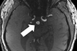
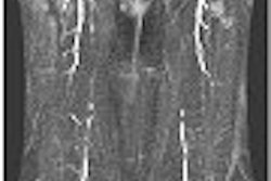
| A 56-year-old woman with a nonhealing ulcer of the right leg had a history of high-dose radiation therapy to her right leg approximately 40 years earlier, following the resection of soft-tissue sarcoma. CTA maximum intensity projection (MIP) with bones removed demonstrates indeterminate density, with calcifications along the course of the posterior tibial artery (arrow). Image demonstrates early filling of the right anterior tibial artery on MRA (right), with no posterior tibial filling and minimal arterial contrast in the left leg. All images courtesy of the Mayo Clinic. |
In addition, CAPR-MRA reached a diagnostic confidence rate of 98% (1,647 of 1.672 segments), better than that of CTA, which achieved a diagnostic confidence rate of 91% (1,531 of 1,672 segments), among the four radiologists reading the images.
For individual vessel segments, CTA achieved greater diagnostic confidence for the popliteal arteries, while there was no difference between CTA and MRA in the tibioperoneal trunks and left proximal anterior tibial artery. In 17 of 22 other segments, there was greater diagnostic conference with MRA results.
High reliability
In conclusion, Mostardi and colleagues determined that although CTA has high reliability and an overall diagnostic confidence of 82%, CAPR-MRA had a superior overall diagnostic confidence of 98%.
"CAPR-MRA can overcome some of the limitations in CTA, in particular those due to calcification or inaccurate bolus timing," she said. "Improvements are notable in distal vessels, which may be important for the clinical planning of bypass [surgery]."
 |
| Subsequent temporal phases of CAPR-MRA demonstrate extensive arteriovenous shunting in the right leg, explaining the early filling not apparent on CTA. There is some distal reconstruction of the posterior tibial artery (arrow), but the proximal segment is occluded. |
Mostardi and colleagues noted that there is some referral bias in the study, because some subjects were recruited for CAPR-MRA after a poor evaluation on CTA. "However, our results indicate that CAPR-MRA can overcome some limitations of CTA and result in improved diagnostic confidence for evaluating lower extremity arteries," according to the group.
The major limitation to vessel evaluation in CAPR-MRA in these subjects, they added, was the loss of signal toward the upper edge of the coil, which resulted in poor assessment of the popliteal arteries in some cases.
Even with those limitations, the Mayo Clinic currently uses CAPR-MRA as its first choice for cases of prior nondiagnostic or limited CTA, known inflow disease or calcification, and imaging of calf vessels.