Researchers at the University of Michigan are taking a new approach to measuring brain activity in patients with Parkinson's disease by using functional MRI (fMRI) to image patients at rest.
The technique deviates from previous studies, which invasively explored brain activity in Parkinson's patients while they were doing certain tasks, rather than resting. Those results helped researchers determine whether or not patients were able to activate task-related areas of their brains or if they were using other compensatory methods to accomplish a task.
"In the past, [patients] have been studied with invasive methods by putting electrodes directly in the part of the brain that is affected by the disease," said study co-author Rachael Seidler, PhD, associate professor in the university's School of Kinesiology and the department of psychology, in an interview with AuntMinnie.com. "These studies were either done in nonhuman animal models or intraoperatively in patients who were going for deep brain stimulation and surgery."
The question for Seidler and her colleagues became: Could they capture the same changes in brain activity while patients were resting using a noninvasive imaging technique? "And, if so," she added, "then it might be useful to track the progression of the disease and evaluate either the current treatment or any future treatment that might be developed to look at their effectiveness in normalizing brain activity."
The researchers published their results in Frontiers in Systems Neuroscience (September 2010, Vol. 4:143, pp. 1-25).
Brain activity
Neural oscillations in a resting state are normal, Seidler said. However, in the part of the brain affected by Parkinson's -- the basal ganglia -- neural oscillations are out of control and spread to other parts of the brain, causing cognition, movement, memory, and other problems.
One of the hallmarks of Parkinson's disease is the loss of the neurotransmitter dopamine in the brain. Dopamine stimulates cognition, memory, and movement and helps neurons connect and communicate in the basal ganglia.
To correct that deficiency, patients receive the drug L-DOPA, which reduces neural oscillations and subsequent disturbances caused by Parkinson's. L-DOPA is a dopamine precursor that can pass through the blood-brain barrier, so the body can use it to manufacture new dopamine.
Functional MRI became the researchers' modality of choice because it can visualize differences in brain oxygenation levels, which reflect metabolic activity in different areas of the brain when they are active.
Deep scans
"It is particularly useful in this case because we can measure the structures that are deep in the brain, such as the basal ganglia," Seidler said. "With some techniques you can only measure some structures at the surface, but with functional MRI you can measure activity in deep brain structures. It has very nice spatial resolution and sufficient temporal resolution to capture the signal that we want."
Functional MRI also can be utilized without a contrast agent, she added.
In the study, the researchers enrolled 25 patients for two fMRI scans. On one day, patients were given L-DOPA. On the second day, they were given a placebo pill. Patients were unaware of what they were given on either day.
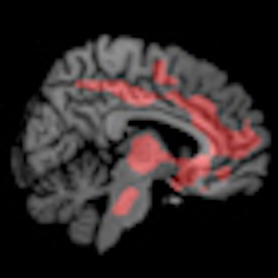
Functional MR images for patients who took the placebo pill and were off the L-DOPA medication indicated a large increase in oscillatory activity in the basal ganglia that spread to other regions of the brain like a ripple effect. When patients were given L-DOPA, their oscillatory brain activity was suppressed, and the brain essentially quieted down.
 |
| Functional MR images of patients who were given placebo pills and were off medication show areas of increased connectivity (red); fMR images of patients who received L-DOPA to suppress brain activity show fewer areas of connectivity (blue). Images courtesy of Rachael Seidler, PhD. |
 |
"The thing that is interesting in both cases, looking at the increased activity when [patients] are off medication and the suppression of activity when they are on medication, [was that] there was a correlation with some behavioral measures in motor and cognitive function," Seidler said. "So, the technique appears to be useful for tracking the disease state, whether they are on treatment are not, and it is doing so in such a way that we can predict their behavior performance, to some extent."
The researchers also found that L-DOPA slowed the neural oscillations too much in some patients. Under those circumstances, fMRI could be helpful in determining the correct concentration of L-DOPA for a patient, they noted.
Seidler and colleagues are following up on this research with studies to determine what additional information can be gained through fMRI. "Eventually, we would like to look at patients at different stages of the disease, including patients in the later stages," she said.
Also, they plan to look at patients who have not yet undergone any treatment to see what fMRI might detect in the early stages of Parkinson's, compared with patients in the later stages.