Cardiac MRI appears to offer prognostic signs of heart failure in patients with arrhythmogenic cardiomyopathy (ACM), according to a study published March 9 in the Journal of the American Heart Association.
The study findings could improve care for patients with ACM, wrote a group led by Dr. Kyeong-Hyeon Chun of Yonsei University School of Medicine in Seoul, Korea.
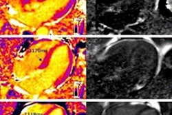
"Cardiac MRI features including late gadolinium enhancement and mapping parameters for left ventricular myocardium can be used in prediction for heart failure events in patients with ACM," the team explained.
Arrhythmogenic cardiomyopathy is a genetic condition in which heart muscle is taken over by scar tissue and fat; it increases the risk of arrhythmias and heart failure. It has tended to be associated with right ventricular changes, but there's more evidence that it can also manifest left ventricular involvement, the authors wrote. They cited a recent autopsy study that found that, in 12% of people who experienced sudden cardiac death and were diagnosed with arrhythmogenic cardiomyopathy, 87% of these showed left ventricular involvement.
"In this context, there have been several reports which investigated the cardiovascular MRI features of the left ventricular phenotype in arrhythmogenic cardiomyopathy and the association with clinical outcomes," it wrote.
Chun's team sought to explore any associations between left ventricular myocardial evaluation using cardiac MRI with clinical outcomes such as heart failure in patients with arrhythmogenic cardiomyopathy via a study that included 60 patients diagnosed with the condition between 2005 and 2020. All patients underwent an electrocardiogram (ECG) and a cardiac MRI exam.
The researchers tracked heart failure-related events -- hospitalization, heart transplant, cardiac death -- as well as ventricular tachycardia events. They also analyzed cardiac MRI findings such as late gadolinium enhancement and native T1, extracellular volume, and T2 mapping values.
Of the 60 patients, 55% had definite arrhythmogenic cardiomyopathy, 13% had borderline ACM, and 32% had possible ACM. Over a median follow-up period of 34 months, 22% of patients had heart failure-related events, and 30% had ventricular tachycardia events.
The team also found the following:
- Only patients with left ventricular late gadolinium enhancement on cardiac MRI had heart failure‐related events.
- When the investigators categorized each mapping parameter by median value, patients with higher native T1, higher T2, and higher extracellular volume experienced more heart failure‐related events.
- There were no significant cardiac MRI differences for ventricular tachycardia events.
These markers on cardiac MRI may prove useful for better assessment of heart failure risk in patients with arrhythmogenic cardiomyopathy, according to Chun and colleagues.
"From the results of our study, we have shown that the presence of late gadolinium enhancement in left ventricular myocardium and higher native T1, ECV, and T2 values measured by cardiac MRI were associated with more heart failure-related events in patients with ACM," they concluded. "Further research is needed to confirm that these left ventricular myocardial characteristics from cardiac MRI are associated with the clinical outcomes in this patient population."