How can a one-time MRI scan of a healthy brain advance the current knowledge on head trauma? Researchers from four institutions plan to find out in a five-year study that will explore how age and gender relate to adverse neurobiological outcomes.
Backed by a $3.6 million grant from the National Institutes of Health (NIH), the study seeks 300 or so participants of various ages and no history of brain injury to undergo gentle mechanical vibrations during an MRI scan. The participants will experience a range of vibrations at modulating frequencies from different angles to view the mechanical behavior of their brains and interactions with the skull. The results will be used to create computer models of normal brain motion under stress to potentially determine which individuals are more susceptible to certain types of trauma at specific times of their lives.
 Philip Bayly, PhD, from Washington University.
"The idea is to characterize, compare, and contrast the [brain's] mechanical behavior and characteristics and develop the tools to create computer models, which could simulate the effects of an automobile crash, an athletic event, or abuse, such as assault," said lead researcher Philip Bayly, PhD, a professor of medical engineering at Washington University in St. Louis. "Essentially, we want the [research] community to make these 'virtual crash dummies' that are anatomically, dynamically, and physically accurate."
Researchers from Washington University; the Henry Jackson Foundation in Bethesda, MD; Johns Hopkins University in Baltimore; and the University of Delaware plan to divide males and females into four age groups: teens from 14 to 17 years, young adults ages 18 to 21, adults from 22 to 50 years of age, and adults older than 50. Previous studies have not included such a range in ages, Bayly said.
The hypothesis is that people of different ages and genders will have varying neurobiological reactions to head trauma. For example, a car crash would have a different impact on a child's brain than an adult's brain.
"The result becomes more subtle when we are tracking a 70-year-old man versus a 40-year-old man, or a 45-year-old man and a 45-year-old woman," Bayly explained to AuntMinnie.com. "All of these differences are potentially important if we are thinking about the mechanism of the injury, what might be the consequences, and how [a physician] would treat it."
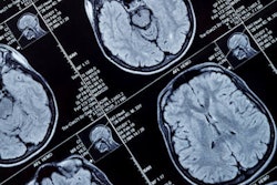
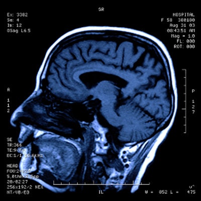
MRI was the modality of choice for this study given its proficiency for imaging brain tissue. Bayly and colleagues plan to use a number of imaging sequences -- including diffusion-tensor imaging (DTI) -- to view and assess the brain's microstructure and white-matter tract integrity. Motion-sensitive MR elastography and tagged MRI also will be utilized to gauge brain and skull reactions.
So what might be the eventual clinical benefits of these computer models on trauma patients? Repeated hits to the head already are associated with an accumulation of tau protein in key regions of the brain and subsequent poor neurological outcomes, such as cognitive impairment, dementia, Alzheimer's disease, and chronic traumatic encephalopathy (CTE). It is very much an open area of research, Bayly said, and the relationship between head trauma and cognitive decline needs to be explored further.
"The question is: Are these changes in the neurobiology of the brain causally related to the changes in cognition and emotion?" he asked. "And if so, at what threshold does tau have to accumulate before there is a change in cognitive function?"
The answers to those questions are "downstream" to the goals of the current endeavor, said Bayly, adding that it is first "very important to understand the upstream, the direct causative, physical phenomena" of what happens when people injure their head or skull.