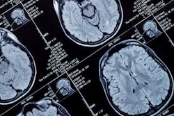
Structural abnormalities visible on the brain MRI and CT scans of infants with the Zika virus (ZIKV) had a significant association with severe clinical outcomes, especially in those initially affected by the virus during the first trimester of pregnancy, according to a study published online July 31 in JAMA Network Open.
Early clinical studies of the Zika virus have shown that it can lead to a wide range of abnormalities affecting the central nervous system, many of which are visible on conventional medical imaging, noted the study authors, led by Drs. Kara-Lee Pool and Kristina Adachi from the University of California, Los Angeles.
In the current study, the researchers from California and Brazil explored the potential association between the presence of abnormalities on the infants' MRI and CT scans at birth and future clinical outcomes.
They evaluated 110 infants exposed to Zika who underwent ultrasound scans followed by either CT and/or 1.5-tesla MRI exams after they were born at a hospital in Rio de Janeiro. In addition to the imaging exams, the infants underwent comprehensive neural, visual, and auditory examinations between March 2016 and June 2017.
Among the Zika-exposed infants, 62% had severe clinical symptoms (prominent central nervous system abnormalities at birth), 5% had moderate symptoms (mild neurologic issues), and 33% had no detected symptoms, the researchers found. The symptoms included neurological conditions (65% of all infants), structural disorders such as microcephaly (49%), fetal brain disruption sequence (45%), and congenital contractures (15%).
In terms of imaging, 65% of the infants had abnormal findings on their brain MRI and CT scans. Roughly 96% of the infants with abnormal MRI and CT scans also had severe clinical symptoms at birth and were most commonly affected by brain calcifications (99% of the infants), brain cortex malformations (95%), ventriculomegaly (93%), and reduced brain volumes (87%), among other brain abnormalities.
The researchers also discovered that certain abnormal findings on MRI and CT were much more common among infants classified as having severe clinical symptoms caused by Zika. For example, all infants with microcephaly, fetal brain disruption sequence, congenital contractures, and poor hearing had abnormal imaging findings, whereas those without these symptoms rarely had abnormal imaging findings.
| Association between Zika-related clinical symptoms and abnormal CT, MRI | |||
| No symptoms | Mild or moderate symptoms | Severe symptoms | |
| % of infants with abnormal CT, MRI | 6% | 17% | 100% |
Furthermore, brain abnormalities were most frequently visible on the MRI and CT scans of infants exposed to Zika in the earliest stages of pregnancy: 63% of infants exposed in the first trimester of pregnancy had neuroimaging abnormalities, compared with 13% of infants in the second trimester and 1% in the third.
The odds ratio for abnormal neuroimaging findings -- notably, malformations in the cortex and brain calcifications -- was almost eight times greater for infants exposed to the virus in the first trimester (p < 0.001).
| Association between time of Zika infection and abnormal CT, MRI | |||
| 1st trimester | 2nd trimester | 3rd trimester | |
| % of infants with any abnormality on CT, MRI | 63% | 13% | 1% |
| % of infants with cortex malformation | 63% | 11% | 2% |
| % of infants with brain calcifications | 64% | 11% | 2% |
"Centered on the findings of Pool, et al, and others, early neuroimaging remains one of the most valuable investigations of the Zika-exposed infant," Dr. Sarah Mulkey, PhD, wrote in an accompanying commentary (JAMA Netw Open, July 31, 2019, Vol. 2:7, pp. e198137).
For most cases, ultrasound should still be the first-line imaging modality, and clinicians should turn to CT and MRI only if the cranial ultrasound scans turn out to be abnormal, Mulkey noted.
"Linking early neuroimaging abnormalities in ZIKV-exposed infants to neurodevelopmental trajectory is the next major important area for understanding the burden of ZIKV on the developing child," she wrote. "Public health and educational systems need to be prepared for the ongoing medical, physical, and educational needs of ZIKV-exposed children."