How low can clinicians go with MRI magnet strength to ensure the safety of patients with implanted cardiac rhythm management (CRM) devices and maintain diagnostic-quality images? The answer is 0.2 tesla, according to an Austrian study published July 4 in the European Journal of Radiology.
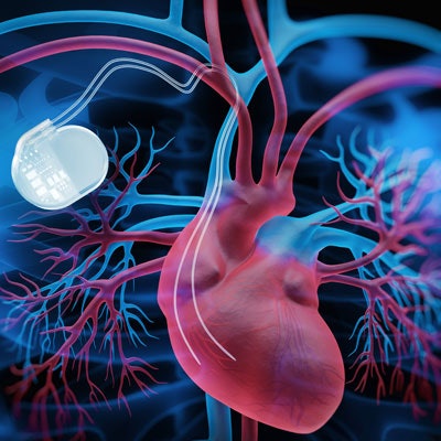
At that magnetic field strength, researchers found that CRM devices -- defined for this study as pacemakers and implantable cardioverter defibrillators (ICDs) -- functioned properly and resulted in no cases of cardiac arrhythmia or other adverse physical effects among the more than 400 patients in the study.
"Both radiologists and cardiologists have to accept that there are still several thousands of patients with non-MRI-conditional CRM systems worldwide. ... For these patients, we absolutely have to provide a safe alternative when standard high-field MRI cannot be performed without concerns," wrote co-authors Dr. Christoph Schukro, PhD, and Stefan Puchner from the Medical University of Vienna. "Taking into consideration our clinical results, low-field MRI is an efficient diagnostic modality for routine scans and provides a high safety regarding clinical and technical complications in this collective."
Clinicians are naturally cautious before performing an MRI scan on patients with CRM devices, given the potentially adverse interactions between the scanner's magnet strength and the device itself -- its leads, battery supply, and general function. While low-field MRI scanners with a magnetic strength of less than 0.5 tesla have proved safe for patients with pacemakers, research is sorely lacking on the safety of patients with ICDs, especially if the devices are not MR conditional, the authors noted.
To fill this research void, Schukro and Puchner looked at four years of routine 0.2-tesla MRI scans (Magnetom Concerto, Siemens Healthineers) on 338 consecutive patients (mean age, 76.1 ± 9.2 years; range, 19-98 years). All the subjects had an implanted CRM device for an average of 4.1 (± 3.2) years. More than one MRI exam was performed in 41 patients (12%), with a maximum of six scans for one patient.
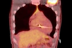
MR images covered a total of 446 different regions of interest, with the most common scans performed on the lumbar spine (206 exams, 46.2%), cerebrum (69, 15.5%), knee (56, 12.6%), and cervical spine (42, 9.4%).
Mean scan time at 0.2 tesla was 34 (± 12) minutes (range, 22-49 minutes), with a specific absorption rate range of 20 to 40 watts/kg. The same electrophysiologist assessed the results of the low-field scans and performance of the CRM devices immediately before and after the scans.
Except for one scan, which was interrupted due to nausea, all 446 MR images were of "good quality and could be interpreted efficiently by the responsible radiologist," the researchers concluded. They found no image artifacts related to the implanted devices. Patients experienced no discomfort, such as a heating sensation, pain, or dizziness, and no cases of arrhythmia or device malfunction.
"Despite the disadvantage of prolonged image acquisition times and the limitation to simple routine MRI scans only (i.e., joints and cranium), this [0.2-tesla MRI] technique reveals some benefits for the clinician," the researchers added. "Besides the comparatively lower overall costs for purchasing and technical maintenance, the open design of low-field MRI scanners enables not only an easier access to the patient for interventional studies but also examinations in special conditions, such as claustrophobic, obese, and pediatric patients."