Using MRI, researchers at Tel Aviv University (TAU) and the University of California, San Francisco (UCSF) believe they have found the reason why certain children with dyslexia are able to overcome their reading disorder and better understand text.
The findings, published online June 14 in PLOS One, target a larger volume of gray matter in the dorsolateral prefrontal cortex of the brain, which is associated with executive functions and working memory.
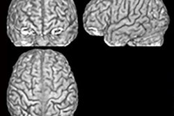
The researchers, led jointly by Dr. Smadar Patael, from TAU's department of communication disorders, and Dr. Fumiko Hoeft, PhD, from UCSF, performed MRI scans on 55 English-speaking children between the ages of 10 and 16 with a wide range of reading abilities. Half of the children had been diagnosed with dyslexia.
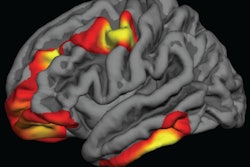
They then crafted a formula to calculate the differences between participants' reading abilities and created maps to illustrate how and where the discrepancies appeared on brain MRI scans.
"We found that the region in the left frontal part of the brain known as left dorsolateral prefrontal cortex was directly related to this discrepancy," Patael said.
To further analyze the mechanics of dyslexia, the researchers followed up with MRI scans on 43 kindergarten students to determine which brain regions helped in overcoming dyslexic deficiencies. Three years later they tested the children's reading abilities.
Their analyses found that the density of neurons in the dorsolateral prefrontal cortex and its association to executive functions and working memory predated mature reading ability and predicted discrepancies in understanding text, regardless of their initial reading abilities.