Researchers at Purdue University are developing a technique based on functional MRI (fMRI) to detect and monitor cerebral vascular disorders and injuries without contrast agents.
The novel fMRI technique tracks an intrinsic blood-related MRI signal that travels with the blood and can be used as a natural biomarker to assess blood flow in a patient. Clinicians could use the signal from symmetric arteries and veins in both hemispheres or the neck to assess cerebrovascular integrity.
"The new method can even be applied on some existing MRI data to calculate the cerebral circulation time," said co-developer Yunjie Tong, PhD, an assistant professor of biomedical engineering, in a release. "This method is safer and noninvasive since we don't inject contrast agents, which can stick to vessels or cause other health problems."
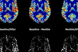
 The technique is based on fMR images that are used to calculate the cerebral circulation time of young, healthy individuals. Image courtesy of Purdue University.
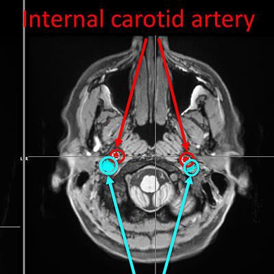
The time delay between the intrinsic signals from the internal carotid artery and the internal jugular vein represents the cerebral circulation time, according to the university. A prolonged time delay would suggest blood flow disturbance in the brain, which could be caused by an abnormality such as a tumor or traumatic brain injury.
The technique was described in a study recently published in the Journal of Cerebral Blood Flow and Metabolism. Tong is looking for vendors, medical institutions, or other collaborators to complete further testing.