Researchers at the University of Chicago Medical Center are reporting early success in using high-powered 7-tesla MRI to visualize and characterize the substructure of multiple sclerosis (MS) lesions. Their hope is to bring the ultrahigh-field magnet to routine clinical practice and expand the options for early diagnosis and treatment of MS.
Preliminary results indicate that 7-tesla MRI provides information not typically seen at 1.5- or 3-tesla imaging for detailed characterization of multiple sclerosis lesions and depiction of their internal structure.
The study was led by Dr. Steffen Sammet, PhD, director of clinical MR physics, who presented the results at the recent American Roentgen Ray Society (ARRS) annual meeting in Washington, DC.
Exploring 7 tesla
In the study, 10 multiple sclerosis patients ranging in age from 33 to 55 years were scanned using 7-tesla MRI (Achieva, Philips Healthcare) and several imaging sequences. Specifically, Sammet and colleagues utilized 2D susceptibility-weighted imaging (SWI), 2D white-matter attenuated (WHAT) inversion recovery turbo spin echo (TSE), and T2-weighted gradient spin echo (GRASE).
Phase images were reconstructed from the SWI data, according to the researchers. All 7-tesla MR images were compared visually and the numbers of lesions seen with each sequence was determined.
Overall, MS lesions had the highest contrast when the WHAT protocol was used, the group found.
"The high spatial resolution and excellent gray-matter/white-matter contrast allowed depicting a number of juxtacortical [adjacent to the cerebral cortex of the brain] lesions and a few gray-matter lesions," Sammet said.
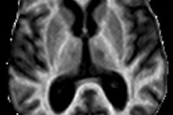
 In this comparison, 7-tesla WHAT (right) shows better lesion contrast and indicates how lesions are associated with small veins, compared with the white matter depicted by 7-tesla GRASE (left). All images courtesy of Dr. Steffen Sammet, PhD.
The WHAT MR images also provided an "excellent depiction of perivascular spaces," Sammet added, and SWI magnitude images illustrated only 93% of the lesions seen on the white-matter attenuated sequence. He credited the difference in performance to "decreased contrast in the weakly proton density-weighted SWI images compared to white-matter attenuated images."
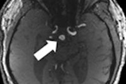
 Some lesions are only seen on 7-tesla SWI phase images (yellow arrows), with lesions appearing as dark rings or with bright centers (red circles) in both SWI magnitude and phase images.
SWI phase images did uncover several interesting features, however. Some lesions were detected on SWI magnitude images but not on the phase images, indicating that contrast in these lesions is due to free water increase, Sammet noted. A free water increase shows as dark spots on MRI and can indicate a disorder such a multiple sclerosis.
Conversely, other MS lesions were seen on phase but not magnitude images. Sammet speculated that the phase-imaged lesions must have significant presence of paramagnetic material, such as iron, which is illuminated on MRI.
Furthermore, a dark outer ring was observed on some lesions on SWI phase images that was not visible in SWI magnitude or white-matter attenuated images. Most lesions visible on SWI magnitude and phase images were associated with venous vasculature.
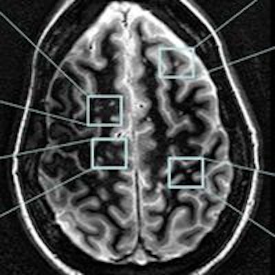
 White-matter attenuated imaging was proficient in depicting MS lesions in various regions of the brain.
Based on the results, Sammet and colleagues were very encouraged that 7-tesla MRI could provide several novel contrast imaging sequences to visualize and characterize multiple sclerosis lesions. It can also differentiate the lesions' substructures not typically visible at 1.5- or 3-tesla MRI.
"Therefore, we have to hope ultrahigh-field MRI of multiple sclerosis might help to better understand the course of the disease and monitor treatment," he added.
Future studies are needed to correlate the various 7-tesla MRI appearances of MS lesions with clinical findings, the researchers noted. In doing so, diagnosis of multiple sclerosis may come sooner and lead to earlier treatment.