Researchers at Case Western Reserve University and University Hospitals Case Medical Center are developing a new imaging method called MR fingerprinting (MRF), which they believe can obtain more information than a traditional MRI.
Preliminary results published online March 13 in Nature indicate that MRF can routinely locate specific cancers as well as multiple sclerosis, heart disease, and other maladies in their early stages when conditions are most treatable.
As explained by co-author Mark Griswold, PhD, a radiology professor at both institutions, body tissue and disease have unique fingerprints that can be used to diagnose clinical conditions. The overall goal is to identify individual tissues and diseases to quantify conditions before they become a problem.
With MRF, researchers simultaneously vary different parts of electromagnetic fields that probe the tissues. The variations make the received signal sensitive to four physical properties that vary from tissue to tissue. These differences are delineated when pattern recognition programs are applied.
The patterns are then charted. Instead of looking at relative measurements from an image, Griswold said quantitative estimates tell one tissue from another. As the technology progresses, these results will determine whether tissue is healthy or diseased and to what degree.
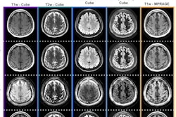
An MRF scan is also much faster than a traditional MRI. MRF scans for different physical properties simultaneously, and researchers have been able to differentiate white matter from gray matter from cerebrospinal fluid in the brain in about 12 seconds, with the promise of performing an exam more quickly in the near future.