While whole-body MRI detected more skeletal lesions on average than conventional imaging among pediatric patients, the modality was less able to find lung metastases in the same group of subjects, according to a study in the February issue of Radiology.
The researchers also found that whole-body MRI was more accurate in the detection of metastatic disease in pediatric patients with solid tumors than in patients with lymphomas.
The study was led by Dr. Marilyn Siegel from the Mallinckrodt Institute of Radiology and Siteman Cancer Center of Washington University School of Medicine (Radiology, Vol. 266:2, pp. 599-609).
An alternative method
Past research has found that the frequency of distant metastasis of tumors in pediatric patients varies according to disease type, from as low as 10% to 12% for sarcomas to as high as 70% for neuroblastomas, the authors noted. As survival rates for children with malignant tumors increase, the overall effect of long-term radiation exposure from medical imaging has become more of a concern.
"Thus, alternative imaging methods that do not use ionizing radiation but allow similar diagnostic accuracy would be particularly attractive," the authors wrote. "Because of its inherently high contrast resolution, MRI is the most important alternative imaging method."
The prospective study recruited 109 male and 79 female subjects from 20 sites (mean age, 10.2 years; range, 1 to 21 years). The 188 individuals were newly diagnosed with lymphoma, neuroblastoma, or soft-tissue sarcoma.
The subjects had previously received CT, conventional MRI, metaiodobenzylguanidine (MIBG) or gallium scintigraphy, or FDG-PET scans. Patients were also eligible for the study if they had undergone imaging within two months of a biopsy or surgical procedure and had a confirmed diagnosis.
Exclusion criteria included contraindications for MRI, such as a cardiac pacemaker or intracranial vascular clip; nonprotocol tumors determined at pathologic examination; previous malignant tumors; being pregnant or nursing; and claustrophobia.
Of the 134 patients who met the study criteria and had available reference standard information, 49 were positive and 85 were negative for metastasis. From that total, 33 patients with positive diagnoses and 33 with negative diagnoses were included in the central reader study.
All patients underwent noncontrast-enhanced whole-body MRI with fast turbo short-tau inversion recovery (STIR) sequences, as well as conventional staging examinations.
Patients with lymphomas received CT of the neck, chest, abdomen, and pelvis, and either gallium scintigraphy or FDG-PET. Those with sarcomas received chest CT, CT or MRI of the primary site, and bone scintigraphy. Patients with neuroblastoma received CT or MRI of the primary site, abdominal and pelvic CT or MRI, and bone scintigraphy or MIBG scintigraphy.
All studies were performed within 21 days of each other before treatment and within two months of any diagnostic or surgical procedure. To minimize recall bias, the researchers required a period of at least one month between interpretation of whole-body MRI studies and those of conventional CT and/or MRI studies for the same case.
Ten pairs of readers reviewed all images, and an independent panel verified the presence or absence of distant metastases.
Image analysis
Among the 33 patients who were negative for metastasis, there were no cases in which a majority of readers recorded a false-positive diagnosis with whole-body MRI. Disease was understaged with a false-negative diagnosis by a majority of readers in eight cases (24%) with whole-body MRI. Of those eight patients, five had lymphoma, two had neuroblastoma, and one had sarcoma.
On a per-patient basis, distant disease was missed by whole-body MRI in the lung in four (50%) of eight patients, in the liver in two (25%), and in distant node (axillary) and tonsil in one (12%) patient each.
Lesions were approximately 1 cm or less in diameter in all of the patients in whom metastasis was missed except for the patients with metastasis in the tonsil axillary node.
 |
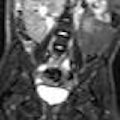
| Coronal whole-body MR images show metastatic neuroblastomas. Image at left of a 4-year-old girl with right adrenal primary tumor shows multiple foci of increased signal intensity in iliac bones and proximal femora. Skeletal and iodine-123 MIBG scintigraphy (not shown) results were negative. Image at right of a 3-year-old boy with left adrenal primary tumor shows diffusely increased signal intensity in both femora. Skeletal and iodine-123 MIBG scintigraphy results (not shown) were positive. Images courtesy of Radiology. |
A per-case analysis among 19 of the 33 patients who were positive for distant skeletal metastases found that, on average, more anatomic regions of skeletal disease were detected with whole-body MRI (5.9) than conventional imaging (3.7). Conventional imaging included scintigraphy for 17 patients and FDG-PET in two cases.
There were no false-negative diagnoses based on missed skeletal lesions at whole-body MRI. Among the 25 patients who were correctly diagnosed for metastasis, there were two patients (8%) in whom most of the readers identified a lesion of less than 1 cm in the lung, and no patients in whom a majority identified a lesion less than 1 cm in the liver.
While previous studies have shown the efficacy of whole-body MRI for skeletal and extraskeletal metastases, the current study "differed in that it was a large, prospective multi-institutional study that used uniform techniques to compare the performance of whole-body MR imaging and conventional imaging for staging of common tumors in pediatric patients," Siegel and colleagues wrote.
The authors reiterated the finding that whole-body MRI was "significantly more accurate for the detection of distant metastatic deposits in solid tumors than in the detection of lymphomas." However, given the results, whole-body MRI "cannot be justified as the study of choice for detection of distant disease in pediatric patients with common malignant tumors, especially those tumors that typically spread to lung and solid organs."