Functional MRI (fMRI) showed changes in brain activation when people with mild traumatic brain injury performed memory tasks, hinting that such injuries could have longer-lasting effects than previously thought, according to a study published in the September issue of Radiology.
Researchers observed differences at fMRI between the injured patients and healthy individuals, even with similar behavioral performances among the two groups. The study suggests that fMRI may have increased sensitivity for assessing mild traumatic brain injury when compared to neuropsychological evaluation alone.
The study was led by Dr. Chi-Jen Chen from the department of radiology at Taipei Medical University in Taiwan (Radiology, September 2012, Vol. 264:3, pp. 844-851).
Not so temporary
The U.S. Department of Health and Human Services estimates that approximately 2 million people suffer nonfatal traumatic brain injury each year, with more than 75% of those cases classified as mild traumatic brain injuries.
Previous research considered mild traumatic brain injury as a temporary affliction, but additional studies have found a person's reduced function can be due to structural changes in the brain from the incident. In addition, results from neuropsychological tests, CT, or MRI are often normal in MTBI patients.
Other fMRI studies on healthy subjects have revealed that increased working memory load is associated with increased activation of the bilateral frontal and parietal regions, a circuitry overlap with regions vulnerable to damage in TBI.
Chen and colleagues' prospective study was conducted between April and September 2010 and included patients at least 17 years of age who had a diagnosis of mild traumatic brain injury. The study excluded patients with a history of epilepsy, cerebrovascular disease, mental retardation, neurodegenerative disorders, prior traumatic brain injury, current use of psychoactive medications, or dental implants that might adversely affect functional MR images, among other factors.
A total of 20 patients with mild traumatic brain injuries were included in the study. Of this group, 10 patients were in motor vehicle collisions, seven had fallen, and three were hit by foreign objects. In addition, a group of healthy volunteers was recruited from a staff of hospital coworkers. These 18 participants were also screened for neurologic, medical, and psychiatric illness.
All 38 participants were scanned on a 3-tesla MRI system (Discovery MR750, GE Healthcare) with an eight-channel head coil. Nine (45%) of the 25 mild traumatic brain injury patients received a follow-up fMRI six weeks after their initial scans.
Memory tasks
The subjects were given a digit span and continuous performance test (CPT) outside the MRI unit prior to a numbers memory task. The digital span is a short-term memory test of how many numbers a participant can remember in sequence, according to the authors. The CPT measures a person's sustained and selective attention and impulsivity.
For example, participants were given a series of numbers and were asked to respond whenever a number shown matched the previous number, the one shown two numbers ago, or the one shown three numbers ago. The "one-back" number is considered a minimal working memory load, while the "two-back" and "three-back" tasks have a moderate and a high working memory load. Each memory test lasted for 30 seconds, during which researchers presented the numbers in sequence.
Chen's team found that the working memory test scores did not significantly differ between the control and MTBI groups, with p-values of 0.90, 0.11, and 0.39 for the one-back, two-back, and three-back tests, respectively.
However, the groups did produce different brain activation patterns as visualized on fMRI scans in response to increasing working memory loads, the authors noted. There was greater activation in control subjects than in the MTBI patients, they wrote.
 |
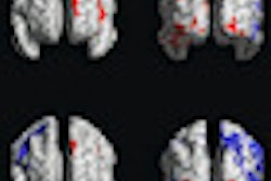
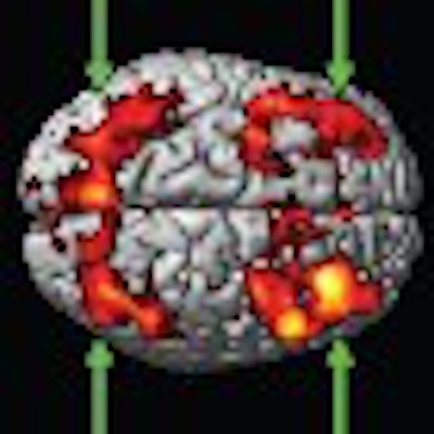
| Functional MRI shows activation maps of one-, two-, and three-back results upon initial scan and at six-week follow-up. Improvement in the working memory circuitry was noted in the follow-up study, which was greater during two-back > one-back conditions (arrows), but less notable with three-back > two-back conditions. All images courtesy of Radiology. |
While the control subjects were able to increase the activity of their working memory circuitry to perform the numbers tasks, the mild traumatic brain injury patients found it more difficult to do so with the moderate and high working load conditions.
The group also noted, however, that the mild traumatic brain injury patients exhibited more activity in some areas outside and inside the working memory circuitry compared to the control subjects. Furthermore, in the six-week follow-up study, MTBI patients showed improved activation in response to greater working memory loads.
Chen and colleagues concluded that fMRI showed differences in brain activation patterns between mild traumatic brain injury patients and control subjects, even though behavioral performance differences were not observed.
"Understanding the mechanisms underlying MTBI impairment and identifying early the brain areas associated with MTBI is critical," the authors wrote. "In the future, functional MR imaging may be used to evaluate and guide treatment strategies, specifically targeting brain areas involved in brain injury recovery."