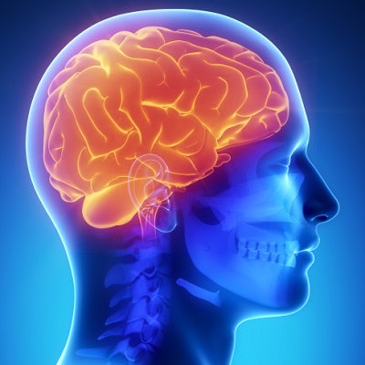
PET imaging results show that long-term depression alters the brain and suggest that different stages of the illness may need different therapies, according to a study published February 26 in Lancet Psychiatry.
People with periods of untreated depression lasting more than a decade had more brain inflammation than those who had fewer than 10 years of untreated disease, wrote a team led by Dr. Jeff Meyer from the Centre for Addiction and Mental Health (CAMH) in Toronto. In earlier research, Meyer and colleagues discovered evidence of inflammation in the brain in patients with clinical depression.
"Greater inflammation in the brain is a common response with degenerative brain diseases as they progress, such as with Alzheimer's disease and Parkinson's disease," he said. "While depression is not considered a degenerative brain disease, the change in inflammation shows that, for those in whom depression persists, it may be progressive and not a static condition."
Meyer and colleagues used PET imaging with an F-18 FEPPA radiotracer to measure brain inflammation in 25 people with more than 10 years of depression, 25 with fewer than 10 years, and 30 people with no depression. They used translocator protein (TSPO) as a marker of inflammation -- F-18 FEPPA has an affinity for TSPO, including increased binding during neuroinflammation.
The researchers found that TSPO levels were 30% higher in the brains of those with long-lasting, untreated depression, compared with those who had shorter periods of untreated disease. The group with long-term depression also had higher TSPO levels than the group with no depression.
Meyer and colleagues are now researching treatment options for later-stage depression, which could include medications that target inflammation, they said.


