A team from Stanford University has used a new molecular imaging agent to visualize a protein critical to cancer cell metabolism, according to research presented June 14 at the virtual Society of Nuclear Medicine and Molecular Imaging (SNMMI) 2021 annual meeting.
The new tracer, called F-18 DASA-23, could help clinicians better assess treatment response in brain cancer patients, the SNMMI said in a statement. The tracer images a "master switch," pyruvate kinase M2 (PKM2), which controls cell metabolism and allows cells to make the building blocks it needs for division.
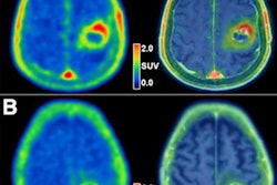
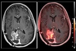
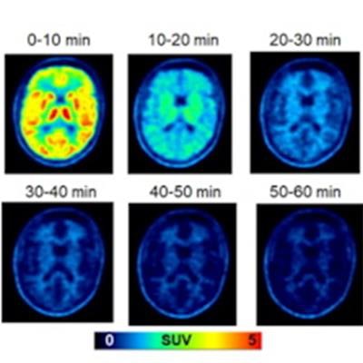
 Clinical evaluation of F-18 DASA-23. (A) Whole-body PET maximum-intensity-projection images at different time points after F-18 DASA-23 administration in a healthy volunteer. (B) Representative axial F-18 DASA-23 PET images of a healthy human brain at various summed time points after tracer administration. (Bettom row) Representative 30-60 minute summed F-18 DASA-23 PET images in patients with intracranial malignancies. Standardized uptake values (SUV) and tumor-to-brain (TBR) values are shown. Images courtesy of SNMMI.
"Until now we've had no way to assess the presence or activity levels of the PKM2 protein involved in that switch," research lead Corinne Beinat, PhD, said in the SNMMI statement. "Through the development of F-18 DASA-23, this is the first time we can noninvasively interrogate the biochemistry of a tumor with respect to this master switch PKM2."
Beinat's group imaged a cohort of both healthy volunteers and glioblastoma patients with PET/MRI using F-18 DASA-23. The group found that the tracer successfully visualized PKM2 in the brain cancer patients and cleared from the bodies of the healthy volunteers.
"This radiopharmaceutical can be very beneficial in assessing whether brain tumor treatments are working," Beinat said. "For example, if a brain tumor is treated with a drug and then imaged with F-18 DASA-23, we can potentially know very quickly whether the therapeutic approach is working. If it's not effective, we won't have to waste more time waiting to see if the tumor itself is shrinking."