A novel PET imaging technique could advance the ability to monitor and guide therapies for patients with type 1 diabetes, Yale University researchers reported in a study published in the August issue of the Journal of Nuclear Medicine.
The PET method is designed to measure beta-cell mass, which includes both functional and nonfunctional beta cells. Low beta-cell function and high signal in the PET scan could indicate dormant beta cells that would respond to a treatment targeting existing cells. A low PET signal would indicate very few viable or dormant beta cells present, however. A patient with that condition might be a candidate for beta-cell transplantation, according to the group led by Jason Bini, PhD, at the Yale University PET Center.
After first screening brain radioligands for their ability to identify beta cells, the researchers chose 12 healthy control subjects and two subjects with type 1 diabetes to receive dynamic PET/CT scans with six tracers (JNM, Vol. 59:8, pp. 1249-1254). They found that carbon-11-(+)-4-propyl-9-hydroxynaphthoxazine (PHNO) was the only radioligand to demonstrate sustained uptake in the pancreas with high contrast, compared with the kidneys, liver, and spleen.
The results provide preliminary evidence that carbon-11-(+)-PHNO is a potential marker of beta-cell mass with binding of dopamine type 3 over dopamine type 2 receptors, according to the researchers.
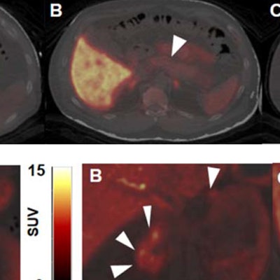
 Top: Representative axial slices of PET/CT images show pancreas uptake (arrowheads) for each dopaminergic radioligand: carbon-11-(+)-PHNO (A), carbon-11-FLB457 (B), and carbon-11-raclopride (C). Bottom: Representative coronal PET/CT images of carbon-11-(+)-PHNO in pancreas (arrowheads) for healthy control (A), C-peptide deficient type 1 diabetic subject (B), and type 1 diabetic subject with detectable C-peptide (C). Images courtesy of JNM.
Bini and colleagues emphasized that additional research is needed to validate the results, but they suggested that the technique could be useful for differentiating beta-cell mass in healthy individuals versus those with type 1 diabetes mellitus.