Although recent data from the National Prostate Cancer Coalition states that one in six men in the U.S. will get prostate cancer during his lifetime, conventional imaging techniques are limited in their capability to detect, stage, and restage this carcinoma.
Nuclear medicine, through hybrid modalities such as PET/CT and SPECT/CT, has offered the most encouragement for clinicians seeking more accurate tools to find and localize prostate cancer; but finding an appropriate radiotracer for imaging evaluation has been difficult.
Prostate carcinoma imaging with FDG-PET has not demonstrated acceptable clinical levels of diagnostic accuracy. PET studies employing choline as a tracer have shown advantages in imaging early androgen-sensitive tumors, while the use of acetate as a tracer suggests capabilities for imaging aggressive androgen-resistant tumors. However, according to researchers from Emory University in Atlanta, these radiopharmaceuticals -- as well as tracers such as methionine and fluorocholine -- have demonstrated mixed results for clinical evaluation.
"Thus, there is a need for a better radiotracer, with little or no urinary excretion, for the evaluation and staging of patients with prostate carcinoma," they wrote in this month's Journal of Nuclear Medicine (January 2007, Vol. 48:1, pp. 56-63).
The Emory scientists collaborated closely with scientists at Nihon Medi-Physics' research center in Chiba, Japan, to examine the potential of using the radiotracer anti-1-amino-3-F-18-fluorocyclobutane-1-carboxylic acid (anti-F-18-FACBC) to better stage or determine prostate cancer.
Anti-F-18-FACBC is a synthetic amino-acid analog whose uptake by prostate carcinoma cells is likely mediated via the sodium-independent L large-neutral amino acid transport system, according to the Emory researchers.
The Japanese group performed in vitro and in vivo studies of anti-F-18-FACBC on animal models utilizing a microPET P4 scanner (Siemens Medical Solutions, Malvern, PA). The team found the tracer had a high accumulation in cancer cells, small excretion into the bladder, and a low accumulation in areas of inflammation.
"In comparison with F-18 FDG, microPET imaging with anti-F-18-FACBC facilitated the visualization of the prostate cancer tissue of the orthotopic prostate cancer transplantation rats with higher contrast," they wrote (JNM, January 2007, Vol. 48:1, pp.46-55). "This suggests the feasibility of anti-F-18-FACBC as a PET tracer for the diagnosis of human prostate cancer."
The Emory team undertook the study of anti-F-18-FACBC in nine patients with a recent diagnosis of prostate carcinoma and six patients with a suspected recurrence of the cancer. All 15 patients first underwent a CT scan of the abdomen and pelvis (80-120 mA) without oral or intravenous contrast for anatomic correlation and attenuation correction of emission data, according to the authors.
The patients then had a 65-minute dynamic PET/CT of the pelvis after intravenous injection of 300-410 MBq of anti-F-18-FACBC, which was followed by a static scan of the abdomen and pelvis at four minutes per bed position. The scans were performed on either a Discovery LS or DST integrated PET/CT scanner (GE Healthcare, Chalfont St. Giles, U.K.).
The images were prospectively assessed by a nuclear radiologist, and the findings were correlated with pathologic, clinical, biochemical, and imaging follow-up for up to one year after scanning.
Of the nine patients who presented with a recent diagnosis of prostate carcinoma, eight were correlated with either sextant biopsy or histologic examination of the surgically removed prostate.
 |
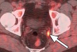
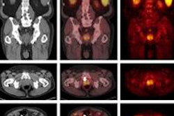
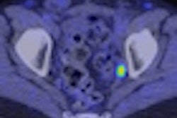
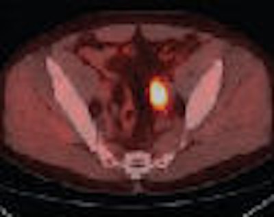
| Axial PET (A) and fused (B) anti-F-18-FACBC images in 67-year-old male patient with intense activity in left external iliac nodes (black arrow in A). SPECT/CT indium-111 capromab-pendetide axial (C) and CT fused (D) images demonstrate no significant activity in this region (white arrow in C). Copyright © by the Society of Nuclear Medicine Inc. Reprinted with permission from “Initial Experience With the Radiotracer Anti-1-Amino-3-18F-Fluorocyclobutane-1-Carboxylic Acid With PET/CT in Prostate Carcinoma,” Schuster DM, John R, Votaw JR, et al, urology department; and F. DuBois Bowman biostatistics department, Emory University in Atlanta, January 2007, Journal of Nuclear Medicine. |
"Visual analysis predicted the presence or absence of carcinoma in 40 of 48 sextants," the authors wrote.
Lymph node status with anti-F-18-FACBC showed seven of the nine patients in concordance with clinical follow-up, with the other two patients noted as indeterminate on imaging. In the six-patient group with suspected recurrence of prostate cancer, the anti-F-18-FACBC scan detected neoplasia in four patients with proven recurrence.
"In two of six patients, mild-to-moderate uptake in the prostate bed resulted in false-positive scans, presumably from the radiation effect noted on biopsy," the authors wrote. "These two patients also had negative anti-F-18-FACBC and conventional imaging of extraprostatic sites."
The researchers observed that anti-F-18-FACBC had relatively intense incidental bowel uptake, as well as occasional low-level uptake in benign inguinal lymph nodes, similar to other radiotracers such as C-11-acetate and F-18 fluorocholine used for PET prostate imaging. They also noted that some pathologically identified benign prostate tissue, including tissue with inflammation, had focal uptake but that it was visually less intense than malignancy.
Bladder activity has limited the utility of FDG and choline-based tracers for prostate imaging, but anti-F-18-FACBC demonstrated relatively little renal excretion and bladder activity.
"Even the most intense bladder activity did not interfere with scan interpretation as has been reported with choline-based PET radiotracers," the authors wrote.
Although the patient cohort was small and heterogeneous, the Emory scientists were encouraged by the results of their pilot study. They believe it is feasible that anti-F-18-FACBC could improve diagnostic imaging of prostate carcinoma both before and after therapy.
"More comprehensive studies must be undertaken to further determine the significance of these findings, to define the optimal time course of imaging, and to find out if comparing early to delayed imaging could increase specificity," they wrote.
By Jonathan S. Batchelor
AuntMinnie.com staff writer
January 18, 2007
Related Reading
Abdominopelvic PET profits from forced diuresis, November 24, 2006
PET/CT locates tumors within prostate but not outside, October 3, 2006
Bone scans not needed in men with low PSA after localized prostate cancer therapy, July 28, 2006
F-18 fluoride PET/CT accurately detects bone metastases in high-risk prostate cancer, March 10, 2006
SPECT/CT pilots prostate brachytherapy in community setting, August 15, 2005
Copyright © 2007 AuntMinnie.com