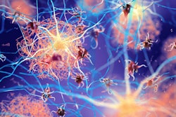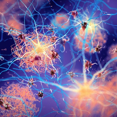
Blood levels of phosphorylated tau (p-tau) in Alzheimer's disease patients appear to be linked more to beta-amyloid plaque deposits in the brain than tau neurofibrillary tangles found on PET imaging, Canadian researchers have found.
The results shed light on an underexplored area of research, wrote a team led by Dr. Pedro Rosa-Neto, PhD, of McGill University in Montreal, Quebec, in a study published December 12 in JAMA Neurology.
"Our findings contribute to the growing understanding of the role of tau phosphorylation in the beta-amyloid cascade and highlight the need for careful interpretation of p-tau biomarkers in [cognitively unimpaired] individuals and as outcomes in disease-modifying clinical trials," the group wrote.
Researchers refer to the complex brain pathology involved in Alzheimer's disease as the amyloid/tau/neurodegeneration framework, or A/T/(N). The concept suggests that neurodegeneration is caused by a cascade of events involving associations between beta-amyloid deposits and tau protein tangles.
PET imaging with radiotracers such as F-18 florbetapir (amyloid PET) and F-18 flortaucipir (tau PET) plays a key role in identifying these affected areas in the brain and are used to identify eligible patients at various stages of Alzheimer's disease for clinical drug trials. Although cerebrospinal fluid (CSF) and plasma concentrations of beta-amyloid and tau protein have also emerged as promising biomarkers for the disease, it remains unclear as to what extent biofluid measurements are associated with beta-amyloid deposits or tau neurofibrillary tangles in the brain, according to the authors.
To elucidate the issue, the researchers evaluated the association between four p-tau cerebrospinal fluid and plasma biomarkers (p-tau181, p-tau217, p-tau231, and p-tau235) with beta-amyloid deposits and tau tangles visualized on patient PET scans. They calculated any associations between PET imaging and fluid biomarkers in 609 patients who were previously enrolled in two large clinical cohorts. Participants included cognitively unimpaired individuals and patients with mild cognitive impairment and dementia.
The investigators' analyses revealed that plasma p-tau181, p-tau217, and p-tau231 had strong associations with amyloid PET findings in patients across the neocortex. Furthermore, p-tau181, p-tau217, and p-tau231 were closely associated with measures of F-18 florbetapir radiotracer uptake by beta-amyloid plaque deposits.
In comparison, associations between plasma concentrations of p-tau181, p-tau217, and p-tau231 with tau PET imaging were lower, including in medial temporal cortices, which is the earliest region to display atrophy over the course of Alzheimer's disease.
Lastly, the group reported that a comparison of these correlations showed that p-tau181, p-tau217, and p-tau231 were significantly more closely associated with amyloid PET than with tau PET, with an 11% difference in plasma p-tau181, a 9% difference in p-tau217, and a 13% difference in p-tau231.
"For all four p-tau phosphorylation sites examined in the cerebrospinal fluid, p-tau was more closely associated with cerebral beta-amyloid plaques than with tau neurofibrillary tangles," Rosa-Neto and colleagues explained.
Taken together with previous research, including autopsy studies, these new findings support evidence of strong associations between beta-amyloid plaques and p-tau biomarkers, which both precede the development of widespread neurofibrillary tangles, the authors added.
"Our study highlights the need for a granular approach to tau biomarkers, in which different tau biomarkers provide complementary but not interchangeable information," they concluded.