PET/CT can help predict which patients have better chances of survival after starting treatment for multiple myeloma, according to a study published March 3 in Blood Advances.
A U.S. team of hematologists and radiologists analyzed FDG-PET/CT scans in newly diagnosed patients before and six months after they began therapy. Results of the imaging indicated which patients responded better and ultimately survived longer, the authors found.
"PET/CT could be incorporated in the post-treatment evaluation of patients with [newly diagnosed multiple myeloma], as it adds crucial prognostic information," wrote senior author Dr. Shaji Kumar, a professor of medicine at the Mayo Clinic in Rochester, MN.
Multiple myeloma is a rare type of blood cancer located primarily in plasma cells in bone marrow, but which can spread throughout the body. Only about half of patients survive more than five years, largely depending on how early the disease is diagnosed. The relative success of treatment -- primarily drug combinations or stem cell transplants -- is determined based on levels of circulating proteins secreted by the diseased plasma cells.
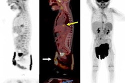
Recent research has shown that whole-body CT, MRI, and PET/CT imaging can detect cancer outside of the bone marrow in these patients, which could be crucial information on whether initial treatments are working.
In this study, Kumar and colleagues, mostly members of the International Myeloma Working Group (IMWG), aimed to confirm this potential value of PET/CT during follow-up exams in patients six months after they began treatment.
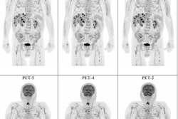
The group analyzed 195 patients who had whole-body PET/CT scans at both diagnosis and approximately six months (range, 2-9 months) after treatment at Mayo Clinic between 2004 and 2020. PET/CT scans were considered negative if areas of increased FDG uptake found at baseline had disappeared, while scans were positive if findings showed new areas of increased FDG metabolism, or if the existing lesions did not entirely disappear.
The end points of the study were time to next treatment (TTNT), measured from the date of diagnosis to any change or intensification of treatment, and overall survival measured from the date of diagnosis to death.
The median follow-up of the entire cohort was 80.6 months. The median age of patients at the time of diagnosis was 61. Fifty (25.6%) patients had negative PET/CT scans at the six-month mark, while 145 (74.6%) had detectable disease, including 30 (15.3%) patients with signs of progression.
The analysis revealed that patients with PET/CT-negative scans had significantly prolonged median TTNT (55.2 vs. 17.8 months) and overall survival (unreached vs. 60.8 months) compared with patients in the PET/CT-positive group.
"A PET/CT [negative scan] at six months confers a significant prognostic advantage for newly diagnosed multiple myeloma patients," the researchers wrote.
Importantly, PET/CT also kept its prognostic significance after researchers adjusted the analysis to include multiple other known risk factors for unfavorable outcomes, the group added.
The authors noted several limitations, namely that due to the nonstandardized reporting of PET/CT results and inconsistency of available standardized uptake values (SUVs), they could not precisely quantify the metabolic activity of the PET/CT scans in the study. As a result, they were not able to objectively compare SUVs between the scans.
Nonetheless, the study highlights the role of PET/CT in the evaluation of multiple myeloma patients in the post-treatment setting, the authors wrote.
"Concurrent assessment of both hematologic and imaging response is relevant in the post-treatment setting for patients with [multiple myeloma], as biochemical information can be crucially improved," the group concluded.