An automated PET image analysis method can track the development of tau protein clumps in the brains of people with Alzheimer's disease, revealing an important clue in how the devastating disease could potentially be treated, according to research published online January 20 in Science Translational Medicine.
After applying their automated PET analysis method to quantify tau progression in patients with Alzheimer's disease at different stages, a group of researchers led by first author Justin Sanchez and senior author Dr. Keith Johnson of Massachusetts General Hospital (MGH) found that tau signal emerged initially in the rhinal cortex -- independently of beta-amyloid levels -- before it spreads in the brain.
As a result, they concluded that treatments that target tau in the rhinal cortex may help delay progression of Alzheimer's disease.
"These findings suggest that [the rhinal cortex] is a biomarker of downstream [tau] spread ... with potential utility for therapeutic trials in which reduction of [tau] spread is an outcome measure," the authors wrote.
Current treatments for Alzheimer's disease have shown reduced efficacy, at least in part because the therapies were administered long after the protein had spread throughout the brain, according to the researchers. A better understanding of where tau pathology originates and how it spreads is needed in order to develop more effective interventions, they said.
Building on recent work that enabled calculation of molecular PET measurements in tau-vulnerable convolutional temporal lobe anatomy, the researchers developed an automated anatomic sampling method to quantify tau signal on PET exams. They then applied their method to PET exams of 443 adult participants from several observational studies of people with Alzheimer's disease. These patients encompassed a wide range of ages, levels of beta-amyloid burden, and degrees of clinical measurements, according to the researchers.
The investigators found that tauopathy initially emerged near the rhinal sulcus in clinically normal people. In a subset of 104 participants who had longitudinal two-year follow-up data, the researchers also observed beta amyloid-associated spread of tau from this site to the nearby neocortex of the temporal lobe, followed by the extratemporal regions. Furthermore, subsequent elevation of tau in the temporal neocortex was associated with age, beta-amyloid, and apolipoprotein E status.
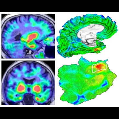
 Four views of the origin of tauopathy in vivo. (Left) Tau PET images for a cognitively normal person. (Top right) 3D rendering of brain surface with tau PET overlay. (Bottom right) Flat map showing topographic detail of surface anatomy with tau origin identified in white outline. Images and caption courtesy of Justin Sanchez and Dr. Keith Johnson of MGH.
What's more, they determined that a greater rate of tau spread was associated with baseline measures of both global beta-amyloid burden and median temporal lobe tau levels.
"These findings are consistent with clinicopathological correlation studies of Alzheimer's tauopathy and enable precise tracking of [Alzheimer's disease]-related [tau] progression for natural history studies and prevention therapeutic trials," the authors wrote.