The use of digital PET to measure FDG uptake in the area of the brain devoted to hearing can help diagnose asymmetrical hearing loss and gauge the potential efficacy of a cochlear implant, according to a study published in the March issue of the Journal of Nuclear Medicine.
Researchers found that decreased FDG uptake occurred in the inferior colliculus and primary auditory cortex on the opposite side of the head to the ear with hearing loss. The ability to determine the severity of this imbalance could help direct otolaryngologists choose the best course of action for their patients.
"Prediction of a successful cochlear implant outcome might benefit from improved imaging with fully digital PET/CT systems, as large parts of the auditory system, including small brain nuclei such as the inferior colliculus, can be assessed for preoperative patient characterization," said lead author Dr. Iva Speck, an otorhinolaryngology resident at the University of Freiburg Medical Center in Germany, in a statement.
Digital high-resolution PET has shown that it can better characterize glucose metabolism in small brain stem nuclei, such as the inferior colliculus, which serves as the primary auditory center and could be the source of hearing loss. Thus, the researchers chose digital PET to investigate asymmetric hearing loss by measuring FDG uptake in the inferior colliculus.
For this study, Speck and colleagues looked at 13 patients with asymmetrical hearing loss who came to the facility to determine their need for a cochlear implant. Their presurgical evaluation included digital PET (Vereos PET/CT, Philips Healthcare) with an injection of 205 MBq (± 20 MBq) of FDG. FDG-PET images were used to determine uptake in the left and right inferior colliculus and left and right primary auditory cortex (JNM, March 2020, Vol. 61: 3, pp. 418-422).
Two experienced readers rated glucose metabolism on a seven-point scale, with 0 indicating hearing symmetry between the left and right inferior colliculus and left and right primary auditory cortex. The scale also rated the degree of asymmetry on both the inferior colliculus and primary auditory cortex.
The digital PET images revealed that significantly lower FDG uptake correlated with the side of the head that was opposite to the ear with the hearing deficiency. In other words, reduced uptake FDG in the left inferior colliculus or left primary auditory cortex was indicative of hearing loss in the right ear. This contralateral pattern was evident with the inferior colliculus in 11 patients (84.6%) and with the primary auditory cortex in 12 patients (92.3%).
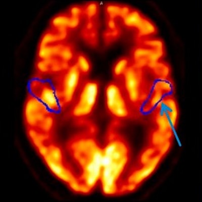
 Axial FDG-PET images at level of primary auditory cortex (top, volume-of-interest outline in blue) and inferior colliculus (bottom) in two representative patients with left-sided (left image) and right-sided (right image) asymmetrical hearing loss. Arrows indicate side of decreased metabolism. Images courtesy of Journal of Nuclear Medicine.
In addition, a longer duration of hearing impairment was associated with a higher metabolism on the contralateral primary auditory cortex. By contrast, duration of hearing impairment did not predict regional glucose metabolism for the ipsilateral primary auditory cortex or either side of the inferior colliculus.
As for the clinical benefits, the findings could help determine which patients with hearing problems are prime candidates for cochlear implants and follow-up therapy.
"Thus, prediction of a successful outcome might benefit from improved imaging with fully digital PET/CT systems, as large parts of the auditory system become accessible for preoperative patient characterization, including small brain stem nuclei such as the inferior colliculus," they concluded. "To confirm these predictions based on presurgical [F-18] FDG PET, further research linking preoperative glucose metabolism with postoperative outcome in patients is underway."