PET imaging with F-18 fluoroethyl-tyrosine (FET) PET can add relevant clinical information for treatment planning and monitoring of children and young adults with brain cancer when MR images are equivocal, according to a study presented at this week's Society of Nuclear Medicine and Molecular Imaging (SNMMI) meeting.
Amino acid PET imaging with FET can be used to evaluate the extent of tumors and help physicians plan for biopsy, surgery, and radiation therapy, as well as track therapy response and tumor recurrence after completion of a treatment cycle.
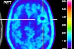
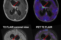
The current study focused on F-18 FET-PET and its diagnostic benefit for imaging pediatric gliomas when conventional MRI cannot provide a clear picture of disease.
In 15 patients with newly diagnosed cerebral lesions suggestive of neoplastic tissue and/or glioma on MRI, FET-PET correctly identified 11 (92%) of 12 lesions as tumors and two (67%) of three lesions as nonneoplastic, for an accuracy rate of 87%. In addition, during the course of the disease, serial FET-PET (17 scans) correctly identified tumor progression/residual tumor in five of six scans and tumor absence in 11 scans, for an accuracy rate of 94%.
"Cancer of the brain and spinal cord are common in children and young adults, and caring for this particular group can be challenging because choice of treatment depends on specific information about the tumor," said lead author Dr. Veronika Dunkl, from the Institute of Neuroscience and Medicine, Forschungszentrum Jülich, in Germany, in a statement.
The study results could significantly improve the clinical management of this patient population, especially when a decision for further treatment is challenging based on conventional MRI alone, she said.